. 2006 Jan;18(1):15-28.
doi: 10.1105/tpc.105.037200. Epub 2005 Dec 2.

Functional diversification of the two C-class MADS box genes OSMADS3 and OSMADS58 in Oryza sativa

Affiliations
Free PMC article

Functional diversification of the two C-class MADS box genes OSMADS3 and OSMADS58 in Oryza sativa

Takahiro Yamaguchi et al. Plant Cell. 2006 Jan.
Free PMC article

Abstract

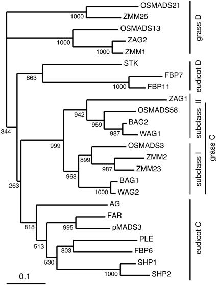
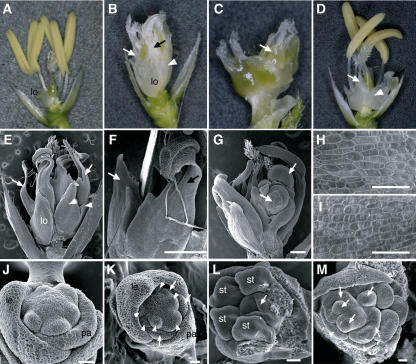
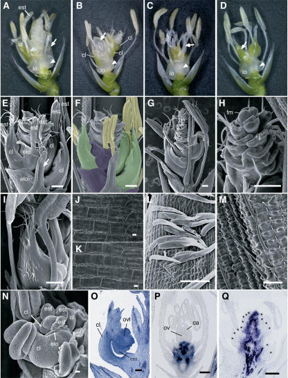
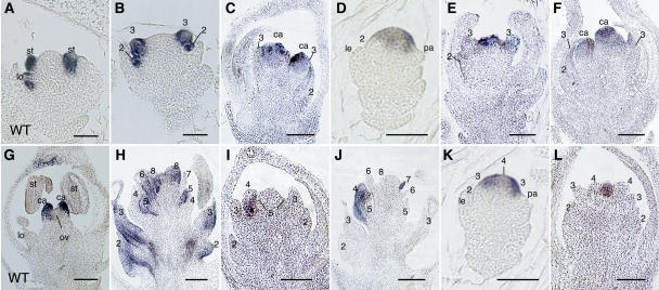
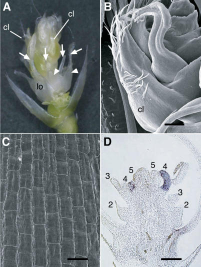
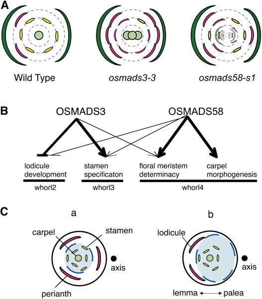
The C-class MADS box gene AGAMOUS (AG) plays crucial roles in Arabidopsis thaliana development by regulating the organ identity of stamens and carpels, the repression of A-class genes, and floral meristem determinacy. To examine the conservation and diversification of C-class gene function in monocots, we analyzed two C-class genes in rice (Oryza sativa), OSMADS3 and OSMADS58, which may have arisen by gene duplication before divergence of rice and maize (Zea mays). A knockout line of OSMADS3, in which the gene is disrupted by T-DNA insertion, shows homeotic transformation of stamens into lodicules and ectopic development of lodicules in the second whorl near the palea where lodicules do not form in the wild type but carpels develop almost normally. By contrast, RNA-silenced lines of OSMADS58 develop astonishing flowers that reiterate a set of floral organs, including lodicules, stamens, and carpel-like organs, suggesting that determinacy of the floral meristem is severely affected. These results suggest that the two C-class genes have been partially subfunctionalized during rice evolution (i.e., the functions regulated by AG have been partially partitioned into two paralogous genes, OSMADS3 and OSMADS58, which were produced by a recent gene duplication event in plant evolution).


Figures

Figure 1.
Figure 1.
Phylogenetic Analysis of MADS Domain Proteins in the AG Subfamily. Analysis was done by the neighbor-joining method, OSMADS21 and ZMM25 were used as outgroups, and the local bootstrap values after 1000 replicates are indicated near the branching points.
Figure 2.
Figure 2.
In Situ Localization of OSMADS3 and OSMADS58 Transcripts in Wild-Type and dl-sup1 Flowers. (A) to (E) In situ localization of OSMADS3 transcripts in wild-type flowers. The stages of flower development proceed from (A) to (E). (F) In situ localization of OSMADS3 transcripts in the dl-sup1 flower. The stage of flower development corresponds to the wild-type flower in (D). (G) to (K) In situ localization of OSMADS58 transcripts in wild-type flowers. The stages of flower development proceed from (G) to (K). (L) In situ localization of OSMADS58 transcripts in the dl-sup1 flower. The stage of flower development corresponds to the wild-type flower in (J). Arrowheads in (A) indicate stamen anlagen where a high level of OSMADS3 transcripts accumulate. Arrows in (A) and (G) indicate the regions that later produce lodicule primordia and that do not accumulate OSMADS3 or OSMADS58 transcripts. Arrows in (F) and (L) indicate ectopic stamens in whorl 4. Lemma and palea primordia in (A) and (G), carpel primordia in (D), and ectopic stamen primordia in (L) are outlined. ca, carpel; le, lemma; lo, lodicule; ov, ovule; pa, palea; st, stamen. Bars = 20 μm.
Figure 3.
Figure 3.
Molecular Characterization of Insertional Mutants of OSMADS3 and RNAi Plants of OSMADS58. (A) Genomic structure of OSMADS3 and position of the T-DNA or Tos17 insertion. Boxes indicate exons, and thick lines indicate introns. (B) Insertion site of Tos17 in osmads3-2. Tos17 sequences are shown in bold. A possible chimeric translation product, terminating within the Tos17 element (asterisk), is shown beginning with amino acid 186. (C) Schematic representation of the transgene construct, which can produce double-stranded RNA of the 3′-half the OSMADS58 sequence with a loop. The positions of the nucleotides of OSMADS58 used in this construct are shown. (D) RT-PCR–based expression analysis of ABC MADS box genes and DL in young flowers of wild-type, osmads3 mutant, osmads58RNAi, and osmads3-2/osmads58RNAi lines. Rice ACT1 was analyzed as a control. The genes subjected to amplification are shown at the left. The number in parentheses indicates the PCR cycle number. Lines from which the RNA samples were isolated are shown at the top. To monitor OSMADS3 expression in osmads3-2, a gene-specific primer and a Tos17-specific primer were used, which resulted in a smaller fragment as compared with samples containing the wild-type allele.
Figure 4.
Figure 4.
Phenotypes of osmads3 Mutant Flowers. (A) Wild-type flower. (B) osmads3-3 flower showing the ectopic development of lodicules in whorl 2 and the homeotic transformation of stamens into lodicules in whorl 3. (C) osmads3-3 flower showing the multiple carpels in whorl 4. For clarity, the organs in whorl 2 and whorl 3 have been removed. Arrow indicates ectopic carpels that formed interior to whorl 4 carpels. (D) osmads3-2 flower. (E) Scanning electron micrograph of the osmads3-3 flower. (F) Scanning electron micrograph of transformed organs in whorl 3 of the osmads3-3 flower. (G) Scanning electron micrograph of ectopic cell masses (arrows) emerging from osmads3-3 carpels. (H) and (I) Scanning electron micrographs showing the epidermal morphology of a wild-type lodicule (H) and a transformed organ in whorl 3 of the osmads3-3 flower (I). (J) and (L) Early development of the wild-type flower. For clarity, some stamens have been removed in (L). (K) and (M) Early development of the osmads3-3 flower. Arrowheads in (B), (D), and (E) indicate ectopic lodicules in whorl 2. White and black arrows in (B) and (D) to (F) indicate complete and incomplete transformation of stamens into lodicules in whorl 3, respectively. Arrows and arrowheads in (K) indicate ectopic lodicules in whorl 2 and in whorl 3, respectively. Arrows in (L) and (M) indicate carpel primordia. Arrowheads in (M) indicate transformed organs in whorl 3. le, lemma; lo, lodicule; pa, palea; st, stamen. Bars = 500 μm in (E) to (G) and 20 μm in (H) to (M).
Figure 5.
Figure 5.
Phenotypes of osmads58RNAi Flowers. (A) osmads58-s1 flower. Arrow indicates a whorl 3 stamen that has been partially transformed into a lodicule. (B) osmads58-s1 flower. Arrow indicates an ectopic cell mass emerging from a carpel-like organ. (C) osmads58-w1 flower. Arrow indicates an ectopic stamen produced interior to the multiple carpels in whorl 4. (D) osmads58-w1 flower. Arrow indicates a carpel-like organ, similar to that formed in osmads58-s1. Arrowheads in (A) to (D) indicate ectopic lodicules in whorl 2. (E) and (F) Scanning electron micrographs of the osmads58-s1 flower showing the reiterative development of lodicules, stamens, and carpel-like organs. Some organs in the outer whorls have been removed to show indeterminate organ development. Arrow in (E) indicates a partially transformed stamen into the lodicule. In (F), each floral organ is indicated by a different color: purple, lodicule; yellow, stamen; green, carpel-like organ. (G) Scanning electron micrograph of the osmads58-s1 flower at anthesis. Some organs in the outer whorls have been removed to show indeterminate organ development. (H) Close-up of (G). The floral meristem (fm), which still produces organs, can be observed. (I) Scanning electron micrograph of a carpel-like organ in osmads58-s1. (J) and (K) Scanning electron micrographs showing the epidermal morphology of a wild-type carpel (J) and the carpel-like organ of the osmads58-s1 flower (K). (L) Scanning electron micrograph of trichomes in the carpel-like organs of the osmads58-s1 flower. (M) Scanning electron micrograph of bristles in wild-type lemma. (N) Scanning electron micrograph of an early stage of the osmads58-s1 flower. (O) Longitudinal section of the carpel-like organ showing an ovule-like structure (ovl) in the osmads58-s1 flower. (P) and (Q) In situ localization of OSH1 transcripts in late stage of the wild-type (P) and osmads58-s1 (Q) flowers. Asterisks in (Q) indicate floral organ primordia. ca, carpel; cl, carpel-like organ; cm, cell mass; elo, ectopic lodicule; est, ectopic stamen developing reiteratively; lo, original lodicule; ov, ovule. Bars = 500 μm in (E) to (I) and 20 μm in (J) to (Q).
Figure 6.
Figure 6.
In Situ Localization of Organ Identity Genes in Wild-Type, osmads3-3, and osmads58-s1 Flowers. (A) and (G) In situ localization of SPW1 (A) and DL (G) transcripts in the wild-type flower. (B) In situ localization of SPW1 transcripts in the osmads3-3 flower. (C) In situ localization of DL transcripts in the osmads3-3 flower. (D) to (F) In situ localization of OSMADS58 transcripts in the osmads3-3 flower. Flower development proceeds from (D) to (F). (H) In situ localization of SPW1 transcripts in the osmads58-s1 flower. (I) and (J) In situ localization of DL transcripts in the osmads58-s1 flower. Flower development proceeds from (I) to (J). (K) and (L) In situ localization of OSMADS3 transcripts in the osmads58-s1 flower. Flower development proceeds from (K) to (L). Whorls are numbered. ca, carpel; le, lemma; lo, lodicule; ov, ovule; pa, palea; st, stamen. Bars = 20 μm.
Figure 7.
Figure 7.
Phenotypes of the osmads3-2/osmads58-s1 Flower. (A) osmads3-2/osmads58-s1 flower. Arrowheads indicate ectopic lodicules in whorl 2. Arrows indicate transformed organs in whorl 3. (B) Scanning electron micrograph of a carpel-like organ developing in the osmads3-2/osmads58-s1 flower. (C) Scanning electron micrograph of epidermal morphology of a carpel-like organ in the osmads3-2/osmads58-s1 flower. (D) In situ localization of DL transcripts in the osmads3-2/osmads58-s1 flower. Whorls are numbered. cl, carpel-like organ; lo, original lodicule in whorl 2. Bars = 20 μm in (B) and (D) and 500 μm in (C).
Figure 8.
Figure 8.
Schematic Representation of the Function of OSMADS3 and OSMADS58. (A) Typical flower phenotypes of the wild type, osmads3, and osmads58. Green, lemma (left) and palea (right); pink, lodicules; yellow, stamens; pale green, carpels or carpel-like organs. (B) Functional diversification of OSMADS3 and OSMADS58. Thin arrows indicate a weak contribution of gene function. (C) The asymmetric arrangement of lodicules and the expression domain of C-class genes. (a) A putative ancestral flower of rice. (b) A present-day rice flower. Blue colors indicate the expression domains of C-class genes.

References

    1. Alvarez-Buylla, E.R., Pelaz, S., Liljegren, S.J., Gold, S.E., Burgeff, C., Ditta, G.S., Ribas de Pouplana, L., Martinez-Castilla, L., and Yanofsky, M.F. (2000). An ancestral MADS-box gene duplication occurred before the divergence of plants and animals. Proc. Natl. Acad. Sci. USA 97 5328–5333. - PMC - PubMed
    1. Ambrose, B.A., Lerner, D.R., Ciceri, P., Padilla, C.M., Yanofsky, M.F., and Schmidt, R.J. (2000). Molecular and genetic analyses of the silky1 gene reveal conservation in floral organ specification between eudicots and monocots. Mol. Cell 5 569–579. - PubMed
    1. Bommert, P., Satoh-Nagasawa, N., Jackson, D., and Hirano, H.-Y. (2005). Genetics and evolution of inflorescence and flower development in grasses. Plant Cell Physiol. 46 69–78. - PubMed
    1. Bowman, J.L., Baum, S.F., Eshed, Y., Putterill, J., and Alvarez, J. (1999). Molecular genetics of gynoecium development in Arabidopsis. Curr. Top. Dev. Biol. 45 155–205. - PubMed
    1. Bowman, J.L., Smyth, D.R., and Meyerowitz, E.M. (1991). Genetic interactions among floral homeotic genes of Arabidopsis. Development 112 1–20. - PubMed

Publication types

MeSH terms

LinkOut - more resources