. 2003 Mar;15(3):655-65.
doi: 10.1105/tpc.009373.

A plant-specific dynamin-related protein forms a ring at the chloroplast division site

Affiliations

A plant-specific dynamin-related protein forms a ring at the chloroplast division site

Shin-ya Miyagishima et al. Plant Cell. 2003 Mar.

Abstract

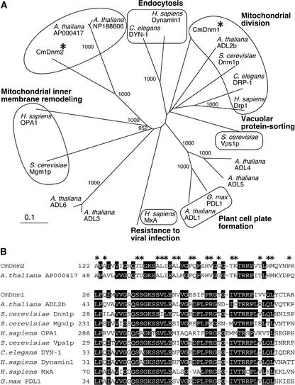
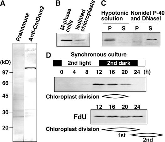
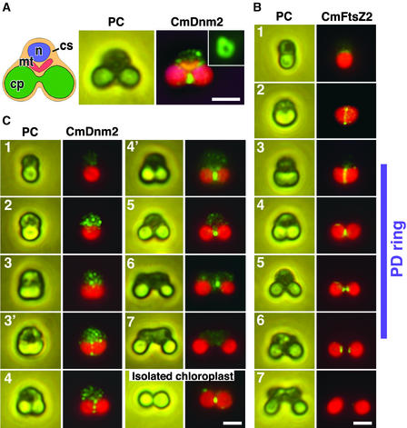
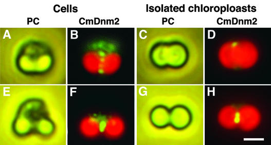
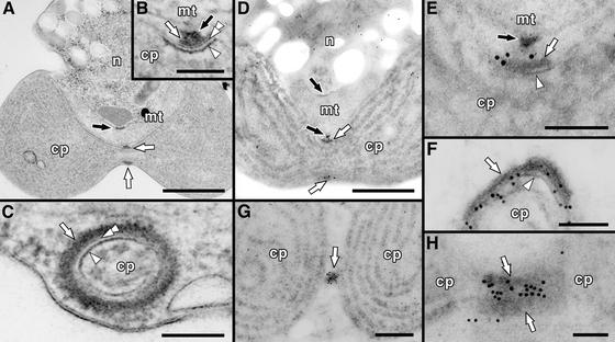
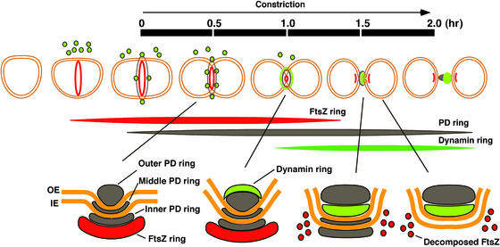
Chloroplasts have retained the bacterial FtsZ for division, whereas mitochondria lack FtsZ except in some lower eukaryotes. Instead, mitochondrial division involves a dynamin-related protein, suggesting that chloroplasts retained the bacterial division system, whereas a dynamin-based system replaced the bacterial system in mitochondria during evolution. In this study, we identified a novel plant-specific group of dynamins from the primitive red alga Cyanidioschyzon merolae. Synchronization of chloroplast division and immunoblot analyses showed that the protein (CmDnm2) associates with the chloroplast only during division. Immunocytochemical analyses showed that CmDnm2 appears in cytoplasmic patches just before chloroplast division and is recruited to the cytosolic side of the chloroplast division site to form a ring in the late stage of division. The ring constricts until division is complete, after which it disappears. These results show that a dynamin-related protein also participates in chloroplast division and that its behavior differs from that of FtsZ and plastid-dividing rings that form before constriction at the site of division. Combined with the results of a recent study of mitochondrial division in Cyanidioschyzon, our findings led us to hypothesize that when first established in lower eukaryotes, mitochondria and chloroplasts divided using a very similar system that included the FtsZ ring, the plastid-dividing/mitochondrion-dividing ring, and the dynamin ring.


Figures

Figure 1.
Figure 1.
CmDnm2 and Two Unknown Arabidopsis Proteins Form a Plant-Specific Monophyletic Group in the Phylogenetic Tree of the Dynamin Family of Proteins. (A) Phylogenetic tree constructed using the N-J method (Saitou and Nei, 1987). Bootstrap values of >900 based on 1000 replications are shown, and some known or predicted functions are annotated according to Hinshaw (2000). Asterisks indicate CmDnm1 and CmDnm2. (B) Comparison of partial amino acid sequences of CmDnm2 and other proteins. Identical amino acids present in more than eight sequences are boxed in black, and asterisks indicate amino acids identical in CmDnm2 and AP000417.
Figure 2.
Figure 2.
CmDnm2 Associates with Chloroplasts Only during the Division Phase. Immunoblot analyses using anti-CmDnm2 antibodies. Twenty micrograms of protein in each sample was separated in each lane, except for in (C), in which samples were obtained from isolated chloroplasts containing 100 μg of protein. (A) Total protein from synchronized M-phase cells was blotted with preimmune antisera or anti-CmDnm2 antibodies. (B) and (C) Dividing chloroplasts were isolated from M-phase synchronous culture (B) and fractionated further into the pellet (P) and supernatant (S) by osmotic bursting or treatment with 0.5% Nonidet P-40 and 0.1 mg/mL DNaseI and then centrifugation (C). (D) Aliquots of the synchronous culture were collected at the indicated times, and total proteins were separated. 5-Flurodeoxyuridine (FdU) was added to the culture at a concentration of 10 μg/mL at 2 h before the onset of the second dark period. The cell cycle was arrested at S-phase by 5-flurodeoxyuridine, whereas chloroplast division occurred continuously, producing four or eight chloroplasts per cell.
Figure 3.
Figure 3.
Immunofluorescence Images of CmDnm2 Accumulating to Form a Ring at the Chloroplast Division Site Only during the Late Stage of Division. (A) Scheme of Cyanidioschyzon cells during M-phase (left), and a phase-contrast image (PC; middle) and an immunofluorescent image (CmDnm2; right) of the same cell showing CmDnm2 localization. When cells were squashed, CmDnm2 ring structures were observed as a closed ring (inset). cp, chloroplast, cs, cytosol; mt, mitochondrion; n, nucleus. (B) Immunofluorescence (CmFtsZ2) and phase-contrast (PC) images of the chloroplast FtsZ ring. The chloroplast division cycle is separated into seven phases. The acorn-shaped chloroplast in the interphase cell (1) changes to a cup (2), trapezoid (3), and dumbbell (4 and 5) shape in M-phase, divides (6), and the resulting chloroplasts are inherited by the daughter cells during cytokinesis (7). The phases in which the PD ring is present are indicated at right. (C) Immunofluorescence (CmDnm2) and phase-contrast (PC) images showing the behavior of CmDnm2 during the chloroplast division cycle and the presence of CmDnm2 on an isolated chloroplast. Green fluorescence indicates CmDnm2 in (A) and (C) and CmFtsZ2 in (B). Red fluorescence indicates the autofluorescence of chlorophyll. Orange fluorescence in (A) shows the mitochondrion stained by MitoTracker. Bars = 2 μm.
Figure 4.
Figure 4.
Immunofluorescence Images Showing That CmDnm2 Binds to Chloroplasts Only during the Late Stage of Division. (A) to (D) Phase-contrast (PC) and immunofluorescence (CmDnm2) images showing the localization of CmDnm2 in a cell ([A] and [B]) or an isolated chloroplast ([C] and [D]) during the early stage of chloroplast division corresponding to phase 4 in Figure 3. (E) to (H) Phase-contrast (PC) and immunofluorescence (CmDnm2) images showing the localization of CmDnm2 in a cell ([E] and [F]) or an isolated chloroplast ([G] and [H]) during the late stage of chloroplast division corresponding to phase 4′ in Figure 3. Bar in (H) = 2 μm for (A) to (H).
Figure 5.
Figure 5.
Electron and Immunoelectron Micrographs Showing the Location of the CmDnm2 Ring. (A) to (C) Electron micrographs of a Cyanidioschyzon cell containing a dividing chloroplast and mitochondrion. (A) A whole cell showing the PD ring and the MD ring. (B) and (C) Magnified cross-sectional (B) and circumferential (C) views of the PD ring showing that it is composed of three rings. (D) to (H) Immunoelectron micrographs showing the localization of CmDnm2. (D) Whole image of a cell containing a chloroplast during the late stage of division, as in (A). (E) Magnified image of the region around the PD ring in (D), as in (B). (F) Circumferential view of the PD ring, as in (C). From its diameter, this ring is thought to be constricted more than the ring shown in (D) and (E). (G) Whole image of a chloroplast at the final stage of division. (H) Magnified image of the region around the PD ring at the final stage of chloroplast division. Gold particles indicate the location of CmDnm2. White arrows, arrowheads, and double arrowheads indicate the outer (at the cytosolic face of the outer envelope), inner (at the stromal face of the inner envelope), and middle (in the intermembrane space) PD rings, respectively. Black arrows indicate the MD ring. cp, chloroplast; mt, mitochondrion; n, nucleus. Bars in (A), (D), and (G) = 500 nm; bar in (B) = 50 nm; bars in (C), (E), (F), and (H) = 100 nm.
Figure 6.
Figure 6.
Scheme of Chloroplast Division by the FtsZ, PD, and Dynamin Rings. Sequential events during chloroplast division are illustrated at top. A cross-section of the division site is shown at bottom. The time scale from trapezoidal chloroplast to just after division (from the start to the end of constriction) (Miyagishima et al., 1999a) is indicated. Red indicates the FtsZ ring; green indicates patches or rings of dynamin-related protein; and black indicates the PD ring (only the cytosolic outer PD ring is shown at top). IE, inner envelope; OE, outer envelope. See text for details.

References

    1. Allen, M.B. (1959). Studies with Cyanidium caldarium, an anomously pigmented chlorophyte. Arch. Microbiol. 32, 270–277. - PubMed
    1. Arimura, S., and Tsutsumi, N. (2002). A dynamin-like protein (ADL2b), rather than FtsZ, is involved in Arabidopsis mitochondrial division. Proc. Natl. Acad. Sci. USA 99, 5727–5731. - PMC - PubMed
    1. Beech, P.L., Nheu, T., Schultz, T., Herbert, S., Lithgow, T., Gilson, P.R., and McFadden, G.I. (2000). Mitochondrial FtsZ in a chromophyte alga. Science 287, 1276–1279. - PubMed
    1. Bi, E., and Lutkenhaus, J. (1991). FtsZ ring structure associated with division in Escherichia coli. Nature 354, 161–164. - PubMed
    1. Bleazard, W., McCaffery, J.M., King, E.J., Bale, S., Mozdy, A., Tieu, Q., Nunnari, J., and Shaw, J.M. (1999). The dynamin-related GTPase Dnm1 regulates mitochondrial fission in yeast. Nat. Cell Biol. 1, 298–304. - PMC - PubMed

Publication types

MeSH terms

Associated data