. 2014 Apr 1;111(13):E1249-55.
doi: 10.1073/pnas.1322134111. Epub 2014 Mar 17.

Embryonic thermosensitive TRPA1 determines transgenerational diapause phenotype of the silkworm, Bombyx mori

Affiliations

Embryonic thermosensitive TRPA1 determines transgenerational diapause phenotype of the silkworm, Bombyx mori

Azusa Sato et al. Proc Natl Acad Sci U S A. .

Abstract

In the bivoltine strain of the silkworm, Bombyx mori, embryonic diapause is induced transgenerationally as a maternal effect. Progeny diapause is determined by the environmental temperature during embryonic development of the mother; however, its molecular mechanisms are largely unknown. Here, we show that the Bombyx TRPA1 ortholog (BmTrpA1) acts as a thermosensitive transient receptor potential (TRP) channel that is activated at temperatures above ∼ 21 °C and affects the induction of diapause in progeny. In addition, we show that embryonic RNAi of BmTrpA1 affects diapause hormone release during pupal-adult development. This study identifying a thermosensitive TRP channel that acts as a molecular switch for a relatively long-term predictive adaptive response by inducing an alternative phenotype to seasonal polyphenism is unique.


Conflict of interest statement

The authors declare no conflict of interest.

Figures

Fig. 1.
Fig. 1.
Embryonic RNAi knockdown of BmTrpA1 induces nondiapause eggs in progeny. (A) Schematic representation of embryonic diapause in the progeny of the silkworm, B. mori. Progeny diapause is determined by environmental temperature during the sensitive period [stages (st.) 20–23] of the mother’s embryonic development. When eggs are incubated at 25 °C, the resultant female moths (25DD) lay pigmented diapause eggs. In contrast, incubation at 15 °C causes the resultant moths (15DD) to lay nondiapause eggs. Nondiapause eggs complete their embryogenesis ∼9 d after oviposition at 25 °C. In contrast, diapause eggs remain in the diapause stage (stage 8). In 25DD pupae, DH released into the hemolymph acts on a G protein-coupled receptor, DH receptor. (B) Effect of dsRNA injection on diapause-egg–inducing activity. Double-stranded RNA of TrpA subfamily genes (trpA1, painless, pyrexia, wtrw, and trpA4) and EGFP were injected into newly laid, nondiapause eggs. Nondiapause egg-inducing activity is represented as the percentage of oviposited eggs in each batch that were nondiapause eggs; trpA1 (D19) and trpA1 (D20–23) indicate BmTrpA1 RNAi-injected larvae that initiated spinning at 19 d and at 20–23 d, respectively. Data represent means ± SD; **P < 0.01 vs. PBS (25DD). (C) Effect of BmTrpA1 dsRNA injection on initiation of spinning. Larval growth rates are represented as days between hatching and initiation of spinning in final- (fifth-) instar larvae. The larvae were checked twice daily at 8:00 AM and 5:00 PM. In B and C, n (egg batch) = 84 (PBS, 25DD); n = 31 (EGFP, 25DD); n = 84 (trpA1, 25DD); n = 81 (others, 25DD); n = 10 (PBS and trpA1, 15DD); and n = 81 (noninjected control, 15DD) in C.
Fig. 2.
Fig. 2.
Developmental expression profiles of BmTrpA1. (A) RT-PCR analysis was performed on 25DD and 15DD during embryonic development. The mRNA levels of BmTrpA1 in 25DD and 15DD (TRPA1, lanes 1–19 and 39–57, respectively) and ActinA3 in 25DD and 15DD (ActA3, lanes 20–38 and lanes 58–76, respectively) were examined. The shaded area indicates the sensitive periods. (B–D) Scanning electron microscope observation of the lateroventral view at stage 20 (B), and lateral view at stages 21 (C) and 23 (D) of embryonic development. The head in C is indicated by red pseudocolor. Each box indicates the body part examined using immunohistochemistry in F, G, and I–K. The arrow indicates yolk granules taken from around the dorsal closure. (Scale bars, 100 µm.) (E) Distribution of BmTrpA1 mRNA in embryos. Whole embryos (E) and yolk (Y) were separated from stage-21 eggs of 25DD and the embryos were dissected to separate the head (H, indicated in C) and thorax and abdomen complex (T+A, indicated in C). Dissected tissues were subjected to RT-PCR analysis of BmTrpA1 (TRPA1, lanes 1–4) and ActA3 (ActA3, lanes 5–8) mRNAs. (F–K) Whole-mount immunohistochemistry was performed using anti-BmTRPA1 antibody at embryo stages 20–23. Immunoreactive signals (red) were detected in the caudal region of stage-20 embryos (F), anal prolegs of stage-23 embryos (I), prolegs of stage 21 embryos (G and H), prolegs of stage-23 embryos (J), and the hump with bristle (socket cell) in stage-23 embryos (K). Arrowheads indicate the immunoreactive signals in each specimen. Asterisks indicate each proleg. The prolegs of stage-21 embryos were stained with DAPI (blue) (H). Magnified images of the tips of prolegs are shown (Insets) (I and J). (Scale bars, 25 µm in F–J and 10 µm in K.)
Fig. 3.
Fig. 3.
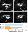
BmTrpA1 knockdown affects immunoreactive intensity of SLb somata in DHPCs. (AD) Representative images of immunohistochemistry of pupal SG using anti-DH antibody. Images are of 15DD (A), and 25DD (B). Also shown are results from pupae injected with BmTrpA1 dsRNA (C) and EGFP dsRNA (D). The dsRNA was injected into newly laid eggs, and the pupal SG was dissected out 4 d after pupation. The arrow indicates the SLb somata. (E) The relative fluorescence intensity in the SLb somata is reported as a percentage of that for 15DD. Each bar represents the mean value of 30 samples ± SD; ***P < 0.001 vs. PBS (25DD). (Scale bar, 100 μm.)
Fig. 4.
Fig. 4.
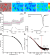
BmTRPA1 is a thermosensitive TRP channel. (A) BmTRPA1-expressing cells were identified using coexpressed DsRed protein (Left). Representative results are shown for Fura-2 Ca2+ imaging at 25.1 °C, 15.4 °C, and 43.7 °C. The pseudocolor indicates intensity of the 340/380-nm fluorescence ratio. (B) Temporal changes in the Fura-2 ratio in BmTRPA1-expressing cells (Upper) and temperature (Lower) in the presence of 2 mM extracellular Ca2+. The average trace (red line) ± SD is shown at the top. Ionomycin was added at 240 s (Iono, gray bar), n = 85. (C) Temporal changes in the current (Upper trace) in a BmTRPA1-expressing cell and temperature (Lower trace) during temperature-fluctuation voltage-clamp whole-cell in patch-clamp analysis. Holding potential was −60 mV throughout the experiment. (D) Representative temperature-response profile for heat-induced BmTRPA1 current, as observed in C. (E) An Arrhenius plot for the heat-induced BmTRPA1 current shows a clear flex point in temperature dependency (data in D were converted). The temperature threshold for BmTRPA1 activation was defined as the point at which the two linear-fitted lines crossed (a flex point). The Q10 value was calculated for each line.
Fig. 5.
Fig. 5.
Chemical compounds activate BmTRPA1 independently of heat, induce diapause, and increase body and cocoon-shell weight. (A) Effect of treatment with chemical compounds on diapause-egg–inducing activity. Eggs were incubated at 15DD with formalin, H2O2, and CA, and the percentage of diapause eggs produced by their progeny was calculated. The moths were divided into two groups: larvae that initiated spinning earlier (<50% of total larvae; early), and larvae that initiated spinning later (>50% of total larvae; late). (B and C) Effects on pupal and cocoon-shell weights. Eggs were incubated at 25 °C or 15 °C in the dark with or without 0.3% formalin. Pupae (B) and cocoon-shell (C) weights after 3 d of pupation are shown. In A–C, 15DD (n = 54), 25DD (n = 141), formalin (0.003%; n = 23, 0.03%; n = 46, 0.3%, n = 87), H2O2 (0.5 mM; n = 64, 5 mM; n = 68, 50 mM; n = 72), CA (0.01 mM; n = 52, 0.1 mM; n = 53, 1 mM; n = 44). Data represent means ± SD; ***P < 0.001, **P < 0.01, *P < 0.05. In A, lowercase letters indicate statistically significant differences in DW-treated vs. chemical-compound-treated animals (a), and early vs. late animals (b). (D–F) Temporal changes in current (Upper trace) in a BmTRPA1-expressing cell, and temperature (Lower trace) during temperature fluctuation without additional chemical compounds (D), with 0.01% formalin (E), and 10 mM H2O2 (F) during cooling in a whole-cell patch-clamp experiment. Membrane potential was held at −60 mV. Note that a sudden increase in current level was observed after H2O2 washout, probably because currents were still gradually developing at that time and were enhanced by the temperature increase.

References

    1. West-Eberhard MJ. Developmental Plasticity and Evolution. New York: Oxford Univ Press; 2003.
    1. Gilbert SF, Epel D. How agents in the environment effect molecular changes in development. In: Gilbert SF, Epel D, editors. Ecological Developmental Biology: Integrating Epigenetics, Medicine, and Evolution. Sunderland, MA: Sinauer Associates; 2009. pp. 38–78.
    1. Mousseau TA, Fox CW. Maternal Effects as Adaptations. New York: Oxford Univ Press; 1998.
    1. Gluckman PD, Hanson MA, Spencer HG. Predictive adaptive responses and human evolution. Trends Ecol Evol. 2005;20(10):527–533. - PubMed
    1. Kostál V. Eco-physiological phases of insect diapause. J Insect Physiol. 2006;52(2):113–127. - PubMed

Publication types

MeSH terms

LinkOut - more resources