. 2004 Jan;74(1):1-10.
doi: 10.1086/380648. Epub 2003 Dec 8.

The breakpoint region of the most common isochromosome, i(17q), in human neoplasia is characterized by a complex genomic architecture with large, palindromic, low-copy repeats

Affiliations

The breakpoint region of the most common isochromosome, i(17q), in human neoplasia is characterized by a complex genomic architecture with large, palindromic, low-copy repeats

Aikaterini Barbouti et al. Am J Hum Genet. 2004 Jan.

Abstract

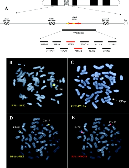
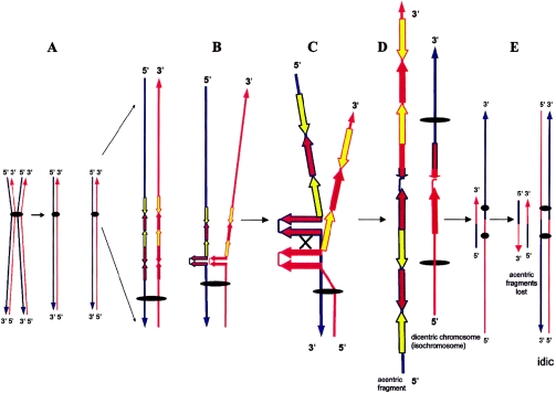
Although a great deal of information has accumulated regarding the mechanisms underlying constitutional DNA rearrangements associated with inherited disorders, virtually nothing is known about the molecular processes involved in acquired neoplasia-associated chromosomal rearrangements. Isochromosome 17q, or "i(17q)," is one of the most common structural abnormalities observed in human neoplasms. We previously identified a breakpoint cluster region for i(17q) formation in 17p11.2 and hypothesized that genome architectural features could be responsible for this clustering. To address this hypothesis, we precisely mapped the i(17q) breakpoints in 11 patients with different hematologic malignancies and determined the genomic structure of the involved region. Our results reveal a complex genomic architecture in the i(17q) breakpoint cluster region, characterized by large ( approximately 38-49-kb), palindromic, low-copy repeats, strongly suggesting that somatic rearrangements are not random events but rather reflect susceptibilities due to the genomic structure.


Figures

Figure  1
Figure 1
i(17q) breakpoint mapping. A, Ideogram of chromosome 17 with a schematic representation of the 17p11.2 genomic region. Thick, horizontal lines at the bottom of the panel represent large insert YAC and BAC clones. The BACs harboring the breakpoints in 10/11 cases are indicated in red. B and C, FISH analysis of patient 2 with the BAC probes RP11-160E2 and CTC-457L16. B, One RP11-160E2 signal is present on the normal chromosome 17 and one RP11-160E2 (yellow) signal of lower intensity is located on the i(17q). C, CTC-457L16 signal (yellow) is present on the normal chromosome 17 but absent on the i(17q). D and E, Dual color hybridization in patient 3, with the adjacent BACs RP11-160E2 (yellow) and RP11-970O14 (red) on the same metaphase. D, One RP11-160E2 signal is located on the normal chromosome 17 and one signal of lower intensity is present on the i(17q). E, Two RP11-970O14 (red) signals, one present on the normal chromosome 17 and one of slightly higher intensity present on the i(17q).
Figure  2
Figure 2
High-resolution physical map of the i(17q) breakpoint cluster region, revealing a complex genome architecture. A, Schematic contig reconstruction of the genomic region based on DNA sequence alignment among completely sequenced clones: BACs CTC-457L16, RP11-160E2, and RP11-135L13; PAC RP1-149D14; fosmids L29232, L29246, L29227, L29233, L29228, L29248, L29225, and L29280; and pieces of unfinished BAC clones, RP11-744A16, CTD-2525F10, and RP11-970O14. The individually sequenced pieces (short blue horizontal bars labeled “1–11”) of RP11-160E2 were used together with sequence information of available fosmid clones (red horizontal lines) to arrive at the final assembly of RP11-160E2. Similarly, sequenced pieces of RP11-744A16 and fosmid sequences were used to reconstruct the genomic region between RP11-160E2 and RP11-135L13. The structure of the sequence assembly of RP11-160E2 is well supported by read pairs from plasmid templates of two sizes spanning the region. We have similar support for RP11-744A16 and all the sequenced fosmids. Each clone/fragment is represented as a horizontal bar and is labeled with a number or clone name. The dashed vertical lines mark the border between different LCR copies. The high DNA sequence identity (>99.8%) and, thus, small differences (1 in 500–1,000 nts) among analyzed copies were identified by distinguishing among cis-morphisms (differences among LCR copies), polymorphisms (differences among libraries), and DNA sequencing errors, each of which is represented at a similar frequency. Yellow and brown arrows refer to REPA and REPB copies, respectively. LCRs are arranged according to their orientation and structure. Note that REPB2 is truncated when compared with REPB1 and REPB3 copies. Both REPA copies contain exons 1–3 of the GRAP gene, and the remaining exons map telomeric to the REPA1. This indicates that REPA2 originated from REPA1; the position of the putative evolutionary breakpoint (“brkpt”) is indicated with a vertical black arrow. Three U3b and two U3a genes map to the arrowhead portion of REPB and REPA, respectively. The red arrowheads of REPAs and REPBs represent a nearly identical ∼4-kb sequence shared among both REPAs and REPBs. B, Restriction enzyme map of the region. The red dashed rectangles represent the REPB copies. Enzyme abbreviations are as follows: K = KpnI; M = MluI; S = SalI; P = PacI. An alternative order (horizontally flipped) of REPB2, REPA2, and REPB3 has been deduced from a previously reported restriction map of the analyzed region (Gao et al. 1997). It is possible that the different order may represent population polymorphism. KYNUR = kynureninase related; SGLTR = Na/glucose-transporter related. C, Dual color fiber-FISH using fosmid clones L292248 (red) and L292227 (green) as probes on stretched control genomic DNA supported the genome architecture proposed by in silico analysis.
Figure  3
Figure 3
Molecular mechanism for i(17q) formation. A, Division of a metaphase chromosome. The double-strand DNA of each chromatid is depicted in red and blue, as is its 3′ and 5′ orientation. B, The formation of an REPB1 and REPB2 palindrome with subsequent breakage (C) and reunion between palindromes on sister chromatids (D) results in the origin of both dicentric and acentric structures. E, Dicentric and acentric structures after replication. The acentric material is lost in dividing cells because of the lack of a centromere. The (iso-)dicentric structure (isochromosome) is retained and will not become disrupted during anaphase because one of the two centromeres is inactivated as a result of their close proximity. Yellow and brown arrows refer to REPA and REPB copies, respectively, and the “×” depicts interchromatid mispairing of direct repeats. According to this model and our present and previously reported data (Fioretos et al. 1999), i(17q) should be formally designated “idic(17)(p11.2).”

References

Electronic-Database Information

    1. Blast 2 Sequences, http://www.ncbi.nlm.nih.gov/blast/bl2seq/bl2.html (for the Blast 2 browser)
    1. GenBank, http://www.ncbi.nih.gov/Genbank/ (for CTC-457L16 [accession number AC003957], RP11-160E2 [accession number AC007952], RP11-744A16 [accession number AC106017], RP11-970O14 [accession number AC036110], CTB-187M2 [accession number AC004448], RP11-1113L8 [accession number AC025627], CTD-2354J3 [accession number AC015935], RP11-311F12 [accession number AC005722], RP11-78O7 [accession number AC015726], RP11-209D14 [accession number AC005730], CTD-2010G8 [accession number AC007963], RP1-149D14 [accession number AJ009617], CTD-2525F10 [accession number AC109313], RP11-135L13 [accession number AC124066], L29232 [accession number AC139083], L29246 [accession number AC139093], L29227 [accession number AC139315], L29233 [accession number AC140106], L29228 [accession number AC144507], L29248 [accession number AC139138], L29225 [accession number AC144506], L29280 [accession number AC139077], mouse RP23-278F12 [accession number AC084044], rat CH230-200D5 [accession number AC105503], and rat CH230-255K6 [accession number AC134746])
    1. Mfold Server, http://www.bioinfo.rpi.edu/applications/mfold/old/rna/form1.cgi
    1. Mitelman Database of Chromosome Aberrations in Cancer, http://cgap.nci.nih.gov/Chromosomes/Mitelman
    1. RepeatMasker Web Server, http://repeatmasker.genome.washington.edu/cgi-bin/RepeatMasker

References

    1. Akgun E, Zahn J, Baumes S, Brown G, Liang F, Romanienko PJ, Lewis S, Jasin M (1997) Palindrome resolution and recombination in the mammalian germ line. Mol Cell Biol 17:5559–5570 - PMC - PubMed
    1. Bailey JA, Gu Z, Clark RA, Reinert K, Samonte RV, Schwartz S, Adams MD, Myers EW, Li PW, Eichler EE (2002) Recent segmental duplications in the human genome. Science 297:1003–100710.1126/science.1072047 - DOI - PubMed
    1. Bailey JA, Liu G, Eichler EE (2003) An Alu transposition model for the origin and expansion of human segmental duplications. Am J Hum Genet 73:823–834 - PMC - PubMed
    1. Barbouti A, Johansson B, Höglund M, Mauritzson N, Strömbeck B, Nilsson PG, Tanke HJ, Hagemeijer A, Mitelman F, Fioretos T (2002) Multicolor COBRA-FISH analysis of chronic myeloid leukemia reveals novel cryptic balanced translocations during disease progression. Genes Chromosomes Cancer 35:127–13710.1002/gcc.10099 - DOI - PubMed
    1. Bi W, Park S-S, Shaw CJ, Withers MA, Patel PI, Lupski JR (2003) Reciprocal crossovers and a positional preference for strand exchange in recombination events resulting in deletion or duplication of chromosome 17p11.2. Am J Hum Genet 73:1302–1315 - PMC - PubMed

Publication types

MeSH terms

Associated data