. 1997 Jul 28;138(2):375-84.
doi: 10.1083/jcb.138.2.375.

The human Arp2/3 complex is composed of evolutionarily conserved subunits and is localized to cellular regions of dynamic actin filament assembly

Affiliations

The human Arp2/3 complex is composed of evolutionarily conserved subunits and is localized to cellular regions of dynamic actin filament assembly

M D Welch et al. J Cell Biol. .

Abstract

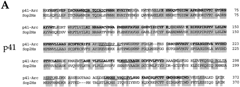
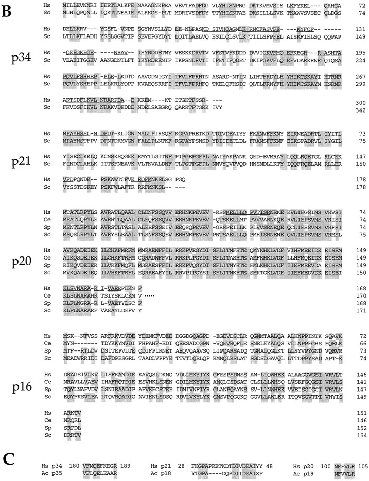
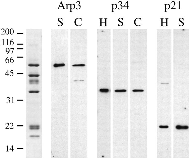
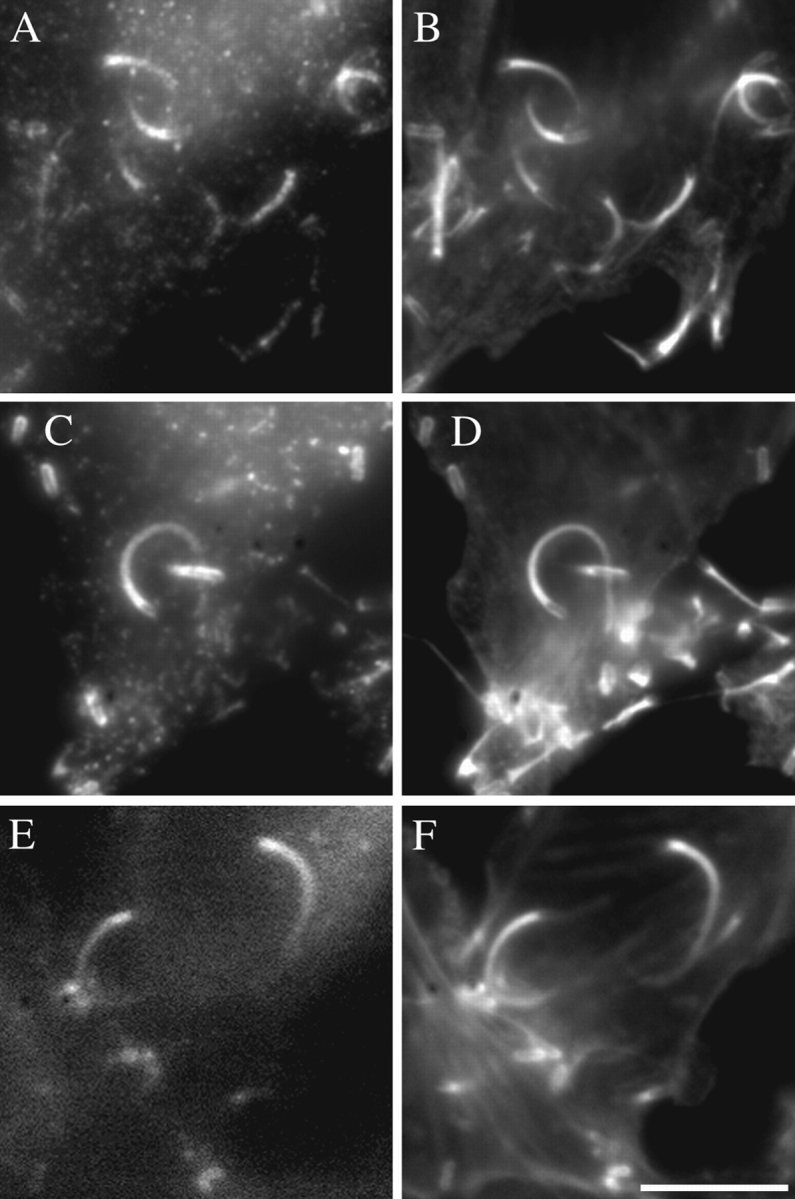
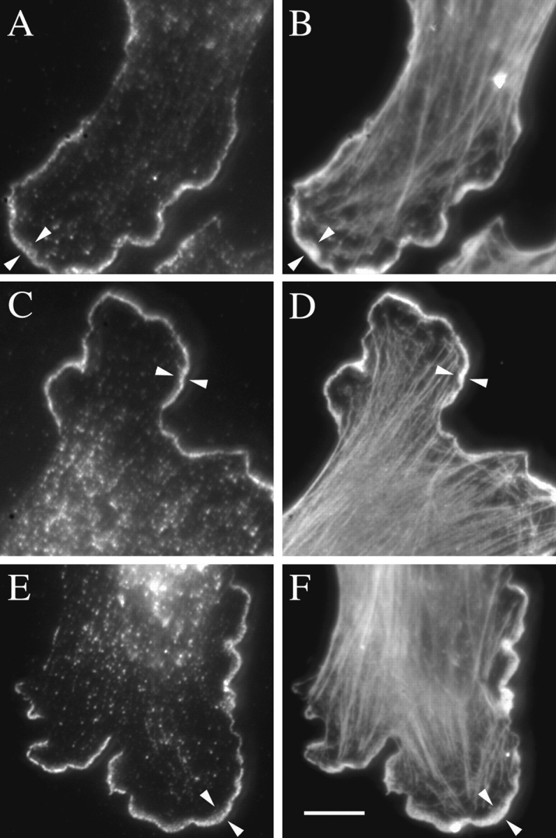
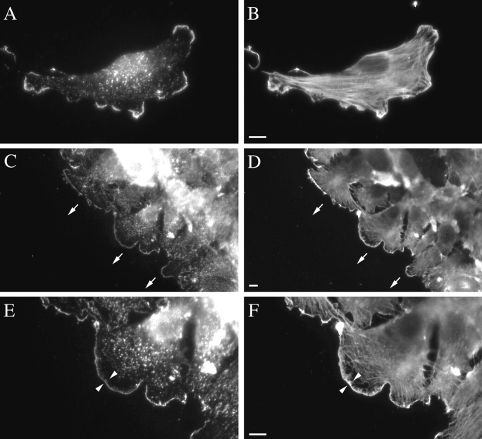
The Arp2/3 protein complex has been implicated in the control of actin polymerization in cells. The human complex consists of seven subunits which include the actin related proteins Arp2 and Arp3, and five others referred to as p41-Arc, p34-Arc, p21-Arc, p20-Arc, and p16-Arc (p omplex). We have determined the predicted amino acid sequence of all seven subunits. Each has homologues in diverse eukaryotes, implying that the structure and function of the complex has been conserved through evolution. Human Arp2 and Arp3 are very similar to family members from other species. p41-Arc is a new member of the Sop2 family of WD (tryptophan and aspartate) repeat-containing proteins and may be posttranslationally modified, suggesting that it may be involved in regulating the activity and/or localization of the complex. p34-Arc, p21-Arc, p20-Arc, and p16-Arc define novel protein families. We sought to evaluate the function of the Arp2/3 complex in cells by determining its intracellular distribution. Arp3, p34-Arc, and p21-Arc were localized to the lamellipodia of stationary and locomoting fibroblasts, as well to Listeria monocytogenes assembled actin tails. They were not detected in cellular bundles of actin filaments. Taken together with the ability of the Arp2/3 complex to induce actin polymerization, these observations suggest that the complex promotes actin assembly in lamellipodia and may participate in lamellipodial protrusion.


Figures

Figure 1
Figure 1
Predicted amino acid sequences of p41-Arc, p34-Arc, p21-Arc, p20-Arc, and p16-Arc aligned to their homologues from diverse organisms. (A) The sequence of p41-Arc is similar to Sop2Hs (these data are available from EMBL/GenBank/DDBJ under accession number Y08999). Bold type indicates the location of the WD repeat sequences and underlined type indicates the location of the peptide sequences used to determine the identity of p41. (B) The sequence of p34-Arc is similar to EMBL/ GenBank/DDBJ accession number Z71650 from Sc. The sequence of p21-Arc is similar to accession number U19103 from Sc. The sequence of p20-Arc is similar to accession numbers U21324 from Ce, Z81317 from Sp, and Z28013 from Sc. The sequence of p16-Arc is similar to accession numbers U88314 from Ce, Z69795 from Sp, and Z38060 from Sc. Underlined type indicates the location of the peptide sequences used to determine the identity of the proteins. (C) Sequences from within p34, p21, and p20 are similar to peptide sequences from the 35-, 18-, and 19-kD subunits of the Ac Arp2/3 complex (Machesky et al., 1994), respectively. Hs, Homo sapiens, Ce, C. elegans, Ac, A. castellanii, Sc, S. cerevisiae, Sp, S. pombe. The sequence data for the subunits of the human Arp2/3 complex are available from EMBL/GenBank/DDBJ under accession numbers AF006084 for p41-Arc, AF006085 for p34-Arc, AF006086 for p21-Arc, AF006087 for p20-Arc, and AF006088 for p16-Arc.
Figure 1
Figure 1
Predicted amino acid sequences of p41-Arc, p34-Arc, p21-Arc, p20-Arc, and p16-Arc aligned to their homologues from diverse organisms. (A) The sequence of p41-Arc is similar to Sop2Hs (these data are available from EMBL/GenBank/DDBJ under accession number Y08999). Bold type indicates the location of the WD repeat sequences and underlined type indicates the location of the peptide sequences used to determine the identity of p41. (B) The sequence of p34-Arc is similar to EMBL/ GenBank/DDBJ accession number Z71650 from Sc. The sequence of p21-Arc is similar to accession number U19103 from Sc. The sequence of p20-Arc is similar to accession numbers U21324 from Ce, Z81317 from Sp, and Z28013 from Sc. The sequence of p16-Arc is similar to accession numbers U88314 from Ce, Z69795 from Sp, and Z38060 from Sc. Underlined type indicates the location of the peptide sequences used to determine the identity of the proteins. (C) Sequences from within p34, p21, and p20 are similar to peptide sequences from the 35-, 18-, and 19-kD subunits of the Ac Arp2/3 complex (Machesky et al., 1994), respectively. Hs, Homo sapiens, Ce, C. elegans, Ac, A. castellanii, Sc, S. cerevisiae, Sp, S. pombe. The sequence data for the subunits of the human Arp2/3 complex are available from EMBL/GenBank/DDBJ under accession numbers AF006084 for p41-Arc, AF006085 for p34-Arc, AF006086 for p21-Arc, AF006087 for p20-Arc, and AF006088 for p16-Arc.
Figure 2
Figure 2
Immunoblots of whole cell extracts probed with anti-Arp3, anti-p34, and anti-p21 antibodies. The far left lane, which shows pure Arp2/3 complex run on a 12% SDS-PAGE gel and stained with Coomassie blue, illustrates the protein composition of the complex. The other lanes are immunoblots of: Swiss 3T3 (S) and chick heart fibroblast (C) extracts probed with anti-Arp3 (Arp3); HeLa (H), Swiss 3T3 (S), and chick heart fibroblast (C) extracts probed with anti-p34 (p34); HeLa (H) and Swiss 3T3 (S) extracts probed with anti-p21 (p21).
Figure 3
Figure 3
Localization of Arp2/3 complex subunits in L. monocytogenes–infected HeLa cells. (A) Distribution of p34-Arc and (B) of actin in the same cell, visualized by immunofluorescence. (C) Distribution of p21-Arc and (D) of actin in the same cell, visualized by immunofluorescence. (E) Distribution of Arp3-EGFP and (F) distribution of actin in the same cell, visualized by staining with rhodamine-phalloidin. Bar, 10 μm.
Figure 4
Figure 4
Localization of Arp2/3 complex subunits in Swiss 3T3 fibroblasts by indirect immunofluorescence. (A) Distribution of Arp3, and (B) of actin in the same cell. (C) Distribution of p34-Arc, and (D) of actin in the same cell. (E) Distribution of p21-Arc and (F) of actin in the same cell. The arrowheads indicate the position of lamellipodia. Bar, 10 μm.
Figure 5
Figure 5
A comparison of the localization of Arp2/3 complex subunits in stationary (A and B) and moving (C–F) Swiss 3T3 fibroblasts by indirect immunofluorescence. (A) Higher magnification view of the distribution of p21-Arc, and (B) of actin in a stationary fibroblast. (C) Lower magnification view of the distribution of p21-Arc and (D) of actin in fibroblasts at the edge of a wound 2 h after wounding a monolayer. Cells move in the direction of the arrows, which point towards the center of the wound. (E) Higher magnification view of p21-Arc and (F) of actin in a cell from C and D, respectively. The arrowheads indicate the position of the lamellipodium. Bars, 10 μm.
Figure 6
Figure 6
Localization of Arp2/3 complex subunits in primary culture fibroblasts from embryonic chick hearts by indirect immunofluorescence. (A) Distribution of Arp3 and (B) of actin in the same cell. (C) Distribution of p34-Arc and (D) of actin in the same cell. The arrowheads indicate the position of the lamellipodia. Bar, 10 μm.

References

    1. Balasubramanian MK, Feoktistova A, McCollum D, Gould KL. Fission yeast Sop2p: a novel and evolutionarily conserved protein that interacts with Arp3p and modulates profilin function. EMBO (Eur Mol Biol Organ) J. 1996;15:6426–6437. - PMC - PubMed
    1. Cossart P. Actin-based bacterial motility. Curr Opin Cell Biol. 1995;7:94–101. - PubMed
    1. Couch FJ, Rommens JM, Neuhausen SL, Belanger C, Dumont M, Abel K, Bell R, Berry S, Bogden R, Cannon-Albright L, et al. Generation of an integrated transcription map of the BRCA2 region on chromosome 13q12-q13. Genomics. 1996;36:86–99. - PubMed
    1. Cramer LP, Mitchison TJ. Myosin is involved in postmitotic cell spreading. J Cell Biol. 1995;131:179–189. - PMC - PubMed
    1. Cramer LP, Siebert M, Mitchison TJ. Identification of novel graded polarity actin filament bundles in locomoting heart fibroblasts: implications for the generation of motile force. J Cell Biol. 1997;136:1287–1305. - PMC - PubMed

Publication types

MeSH terms