. 1998 Oct;72(10):8289-300.
doi: 10.1128/JVI.72.10.8289-8300.1998.

Structure and distribution of endogenous nonecotropic murine leukemia viruses in wild mice

Affiliations

Structure and distribution of endogenous nonecotropic murine leukemia viruses in wild mice

K Tomonaga et al. J Virol. 1998 Oct.

Abstract

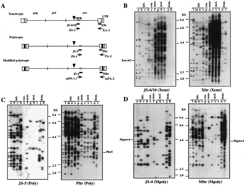
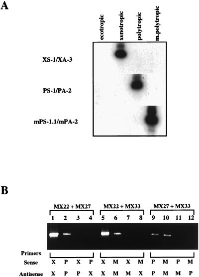
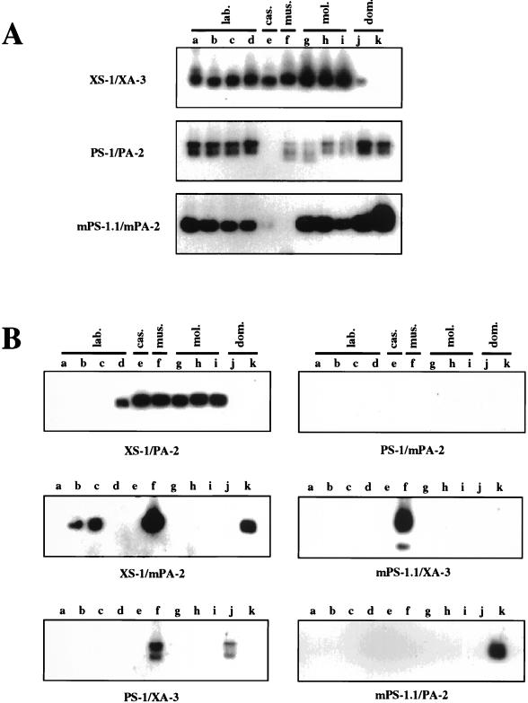
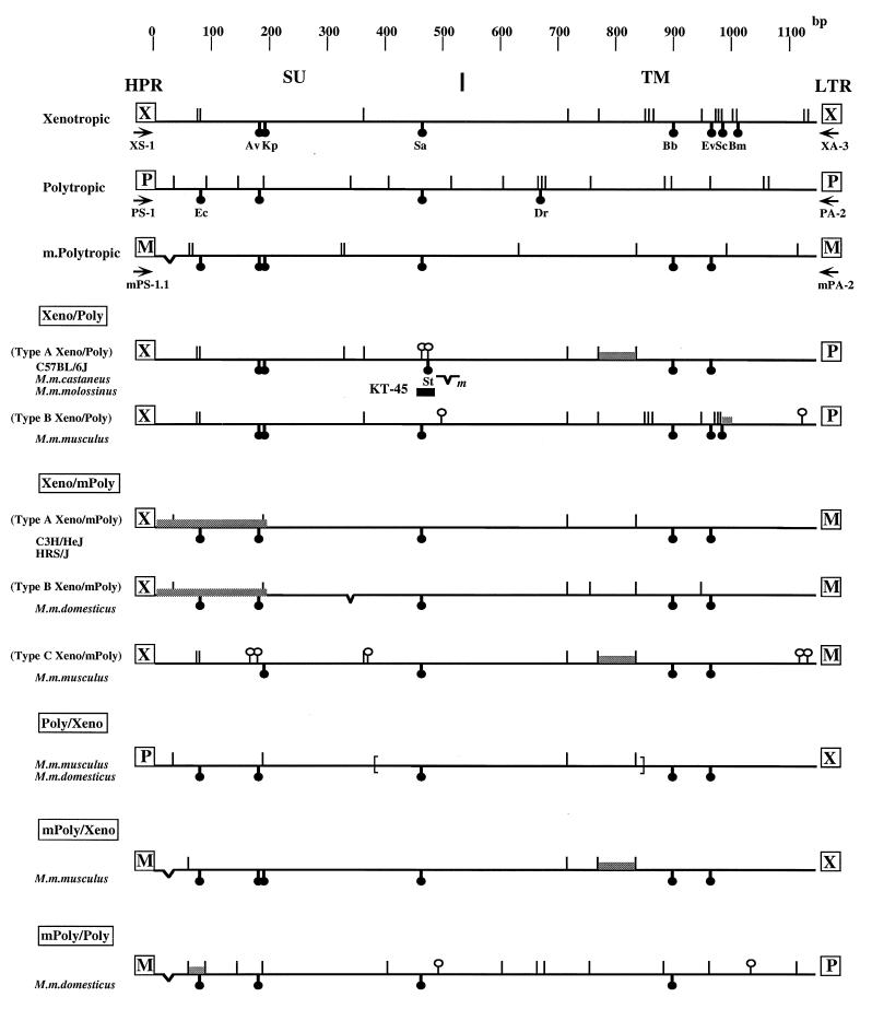
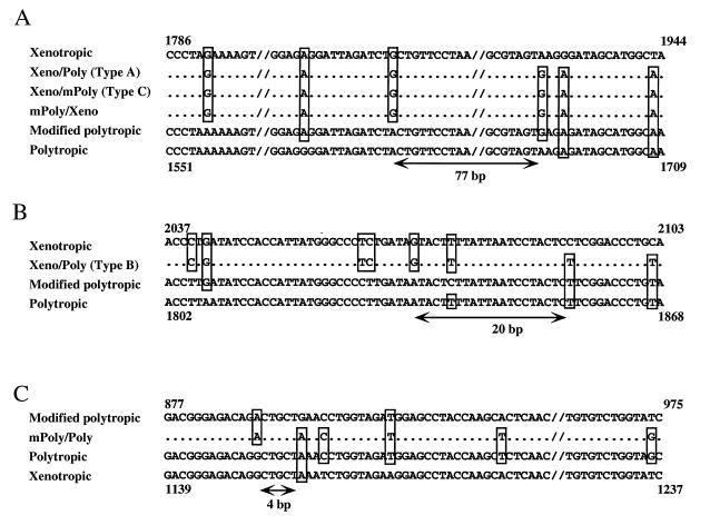
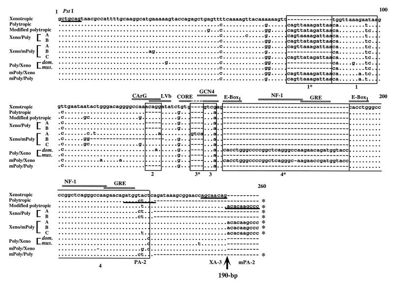
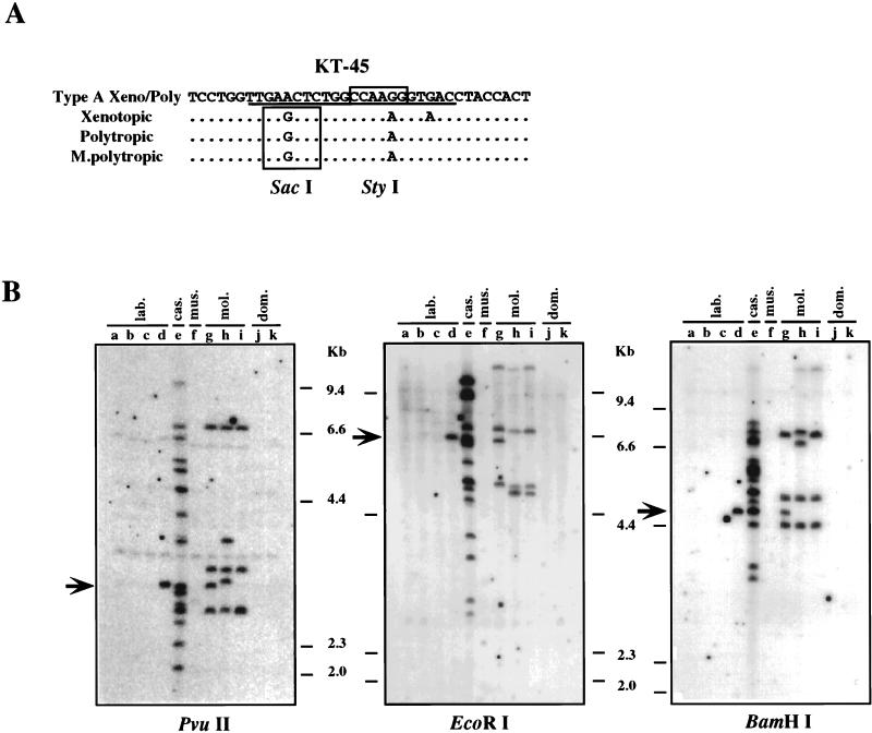
Virtually all of our present understanding of endogenous murine leukemia viruses (MLVs) is based on studies with inbred mice. To develop a better understanding of the interaction between endogenous retroviruses and their hosts, we have carried out a systematic investigation of endogenous nonecotropic MLVs in wild mice. Species studied included four major subspecies of Mus musculus (M. m. castaneus, M. m. musculus, M. m. molossinus, and M. m. domesticus) as well as four common inbred laboratory strains (AKR/J, HRS/J, C3H/HeJ, and C57BL/6J). We determined the detailed distribution of nonecotropic proviruses in the mice by using both env- and long terminal repeat (LTR)-derived oligonucleotide probes specific for the three different groups of endogenous MLVs. The analysis indicated that proviruses that react with all of the specific probes are present in most wild mouse DNAs tested, in numbers varying from 1 or 2 to more than 50. Although in common inbred laboratory strains the linkage of group-specific sequences in env and the LTR of the proviruses is strict, proviruses which combine env and the LTR sequences from different groups were commonly observed in the wild-mouse subspecies. The "recombinant" nonecotropic proviruses in the mouse genomes were amplified by PCR, and their genetic and recombinant natures were determined. These proviruses showed extended genetic variation and provide a valuable probe for study of the evolutionary relationship between MLVs and the murine hosts.


Figures

FIG. 1
FIG. 1
Distribution of nonecotropic proviruses in wild mice. (A) Locations of the group-specific PCR primers and probes. The structures of endogenous nonecotropic proviruses are shown. The approximate positions of the gag, pol, and env genes and the relative sizes of the LTRs are also indicated. The black boxes within the LTRs of the polytropic and modified polytropic viruses corresponded to the 190-bp inserted sequence (24). Arrowheads show conserved PvuII sites in the env gene in nonecotropic provirus genomes. (B to D) Unblotting analysis of PvuII-digested mouse DNAs was performed with xenotropic oligonucleotide probes (JS-6/10, env; Xltr, LTR) (B), polytropic oligonucleotide probes (JS-5, env; Pltr, LTR) (C), and modified polytropic oligonucleotide probes (JS-4, env; Mltr, LTR) (D). Lanes: a and l, AKR/J; b and m, HRS/J; c and n, C3H/HeJ; d and o, C57BL/6J (lab.); e and p, CAST/Ei (M. m. castaneus) (cas.); f and q, CZECH II/Ei (M. m. musculus) (mus.); g and r, MOLC/Rk (M. m. molossinus); h and s, MOLF/Ei (M. m. molossinus); i and t, MOLG/Dn (M. m. molossinus) (mol.); j and u, WSB/Ei (M. m. domesticus); k and v, ZALENDE/Ei (M. m. domesticus) (dom.). Known provirus loci that comigrate with wild-mouse fragments are shown on the side. Boxed bands indicate a comigrating fragment between two different probes. The approximate positions of molecular markers are also shown. Identically sized bands detected with both env and LTR probes are indicated by dots in lanes a and l.
FIG. 2
FIG. 2
Group-specific PCR primers for endogenous nonecotropic proviruses and specificity of the PCRs. (A) Specificity of the primer pairs. A Southern blot analysis of PCRs with each matched primer pair is shown. Each ecotropic (MX14) and nonecotropic (xenotropic, MX22; polytropic, MX27; modified polytropic, MX33) (42) proviral clone was used for the templates of the PCR. Primer pairs for each PCR are indicated on the left. (B) Specificity of the PCR. EtBr staining of the gels is shown. PCRs were performed in the presence of two different clones of nonecotropic proviruses with each combination of the primers. Templates: lanes 1 to 4, MX22 + MX27; lanes 5 to 8, MX22 + MX33; lanes 9 to 12, MX27 + MX33. Primer pair: lanes 1 and 5, XS-1/XA-3; lanes 2 and 9, PS-1/PA-2; lanes 6 and 10, mPS-1.1/mPA-2; lane 3, XS-1/PA-2; lane 4, PS-1/XA-3; lane 7, XS-1/mPA-2; lane 8, mPS-1.1/XA-3; lane 11, PS-1/mPA-2; lane 12, mPS-1.1/PA-2.
FIG. 3
FIG. 3
Detection of endogenous proviruses in genomic DNAs. Each group of nonecotropic proviruses (A) and recombinant forms of nonecotropic proviruses (B) were detected by PCR. The PCR products were analyzed by Southern blot hybridization with a 32P-labeled oligonucleotide probe (see Materials and Methods). The primer pairs used for the PCRs are indicated on the left (A) or at the bottom (B). Lanes: a, AKR/J; b, HRS/J; c, C3H/HeJ; d, C57BL/6J; e, CAST/Ei (M. m. castaneus); f, CZECH II/Ei (M. m. musculus); g, MOLC/Rk (M. m. molossinus); h, MOLF/Ei (M. m. molossinus); i, MOLG/Dn (M. m. molossinus); j, WSB/Ei (M. m. domesticus); k, ZALENDE/Ei (M. m. domesticus).
FIG. 4
FIG. 4
Schematic representation of env structures of recombinant proviruses. The nucleotide sequences of recombinant proviruses are compared with those of each nonecotropic provirus. Sequences of NZB (37) and CWM (32) were used for standard xenotropic viruses and MX27 and MX33 (42) were used for polytropic and modified polytropic viruses, respectively. Symbols: (|) nucleotide differences from consensus nonecotropic provirus sequence; (○|) unique nucleotide differences from consensus nonecotropic provirus sequence in recombinant proviruses; (☻) unique restriction enzyme site: Ec, EcoRII; Av, AvrII; Kp, KpnI; Sa, SacI; St, StyI; Dr, DraI; Ev, EcoRV; Sc, ScaI; Bm, BsmI; (V) deletion. The shaded areas indicate the possible recombinant region in each provirus. The location of the KT-45 hybridization probe is indicated. m in the type A Xeno/Poly recombinant virus indicates the deletion site in the clone from M. m. molossinus. The deletion region in the Poly/Xeno recombinant proviruses are shown by brackets. The boundary of SU and the TM region is also shown at the top. Arrows indicate PCR primers.
FIG. 5
FIG. 5
Possible recombinant regions in recombinant proviruses. (A) Sequences of possible recombination sites of type A Xeno/Poly, type C Xeno/mPoly, and mPoly/Xeno recombinant proviruses. (B and C) Recombination sites of type B Xeno/mPoly (B) and mPoly/Poly (C) recombinant proviruses. Nonecotropic provirus sequences are shown at the top and bottom. Only bases differing from those of at least one nonecotropic provirus are shown. The deduced recombination sites are indicated by arrows and correspond to the region indicated in Fig. 4. Sources of sequence data: xenotropic, NZB (37); polytropic, MX27 (42); modified polytropic, MX33 (42).
FIG. 6
FIG. 6
U3 sequences of recombinant proviruses. The nucleotide sequences of recombinant proviruses are compared with analogous regions of xenotropic (NZB) (37), polytropic (MX27) (42), and modified polytropic (MX33) (42) proviruses. The sequence of the NZB xenotropic virus was used as a standard sequence. Dots indicate nucleotide identity. Dashes indicate the absence of a nucleotide. Direct repeats and unique sequences present in the proviruses are boxed; these regions are designated 1*, 1, 2, 3*, 3, 4*, and 4. Potential enhancer sequence regions are also indicated by the shaded bar. The position of the 190-bp insertion is shown by the arrow. Proviruses with the 190-bp insertion are indicated by asterisks on the 3′ end of the sequences. Primer sequences are underlined. The conserved PstI site is also shown.
FIG. 7
FIG. 7
Distribution of type A Xeno/Poly recombinant provirus. (A) Specific oligonucleotide probe for the type A Xeno/Poly recombinant proviruses. The nucleotide sequence of the type A Xeno/Poly recombinant provirus is compared with analogous regions of xenotropic (NZB) (37), polytropic (MX27) (42), and modified polytropic (MX33) (42) proviruses. The sequence of the specific probe, KT-45, is underlined. Unique restriction recognition sites are boxed. (B) Detection of type A Xeno/Poly recombinant proviruses. Unblotting was performed by using PvuII-, EcoRI-, or BamHI-digested genomic DNAs. Lanes: a, AKR/J; b, HRS/J; c, C3H/HeJ; d, C57BL/6J; e, CAST/Ei (M. m. castaneus); f, CZECH II/Ei (M. m. musculus); g, MOLC/Rk (M. m. molossinus); h, MOLF/Ei (M. m. molossinus); i, MOLG/Dn (M. m. molossinus); j, WSB/Ei (M. m. domesticus); k, ZALENDE/Ei (M. m. domesticus). The approximate positions of molecular markers are shown on the right. Arrows indicate comigrating bands among different subspecies of mice.

References

    1. Battini J-L, Heard J M, Danos O. Receptor choice determinants in the envelope glycoproteins of amphotropic, xenotropic, and polytropic murine leukemia viruses. J Virol. 1992;66:1468–1475. - PMC - PubMed
    1. Boeke J D, Stoye J P. Retrotransposons, endogenous retroviruses, and the evolution of retroelements. In: Coffin J M, Hughes S H, Varmus H E, editors. Retroviruses. Cold Spring Harbor, N.Y: Cold Spring Harbor Laboratory; 1997. pp. 343–435. - PubMed
    1. Bowes C, Li T, Frankel W N, Danciger M, Coffin J M, Applebury M L, Farber D B. Localization of a retroviral element within the rd gene coding for the b-subunit of cGMP-phosphodiesterase. Proc Natl Acad Sci USA. 1993;90:2955–2959. - PMC - PubMed
    1. Ch’ang L-Y, Yank W K, Myer F E, Koh C K, Boone L R. Specific sequence deletions in two classes of murine leukemia virus-related proviruses in the mouse genome. Virology. 1989;168:245–255. - PubMed
    1. Chattopadhyay S K, Lander M R, Gupta S, Rands E, Lowy D R. Origin of mink cytopathic focus-forming (MCF) viruses: comparison with ecotropic and xenotropic murine leukemia virus genomes. Virology. 1981;113:465–483. - PubMed

Publication types

LinkOut - more resources