. 1998 Jul 7;95(14):8181-6.
doi: 10.1073/pnas.95.14.8181.

Mutations in the Delta7-sterol reductase gene in patients with the Smith-Lemli-Opitz syndrome

Affiliations
Free PMC article

Mutations in the Delta7-sterol reductase gene in patients with the Smith-Lemli-Opitz syndrome

B U Fitzky et al. Proc Natl Acad Sci U S A. .
Free PMC article

Abstract

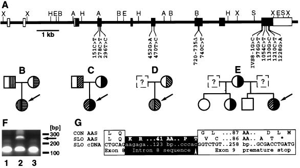
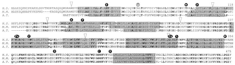
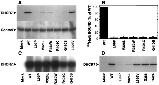
The Smith-Lemli-Opitz syndrome (SLOS) is an inborn disorder of sterol metabolism with characteristic congenital malformations and dysmorphias. All patients suffer from mental retardation. Here we identify the SLOS gene as a Delta7-sterol reductase (DHCR7, EC 1.3.1. 21) required for the de novo biosynthesis of cholesterol. The human and murine genes were characterized and assigned to syntenic regions on chromosomes 11q13 and 7F5 by fluorescense in situ hybridization. Among the mutations found in patients with the SLOS, are missense (P51S, T93M, L99P, L157P, A247V, V326L, R352W, C380S, R404C, and G410S), nonsense (W151X), and splice site (IVS8-1G>C) mutations as well as an out of frame deletion (720-735 del). The missense mutations L99P, V326L, R352W, R404C, and G410S reduced heterologous protein expression by >90%. Our results strongly suggest that defects in the DHCR7 gene cause the SLOS.


Figures

Figure 1
Figure 1
Mutations in the DHCR7 gene of patients with the SLOS. (A) Structure of the gene. Noncoding (open boxes) and coding exons (filled boxes) are shown. Restriction enzyme cleavage sites (A, AccI; B, BamHI; H, HindIII; S, SpeI; X, XhoI) and mutations are indicated. (B-E) Families with SLOS. The index patient is indicated by the arrow. Heterozygotes and homozygotes for mutations in the DHCR7 gene are shown. (B) Patient SLO8; mutations R404C (▥) and A247V (▤). (C) Patient SLO1; mutations V326L (▨) and W151X (▪). (D) Patient SLO6; mutations P51S (▩) and IVS8–1G>C (▧). (E) patient SLO2; mutations V326L (▨) and W151X (▪). (F) Abnormal reverse transcription-PCR-product (arrow) from fibroblasts from patient SLO5 (lane 2) not seen in cDNA from a control person (lane 1) and patient SLO9 (lane 3). Oligonucleotides c898s (GACCACTTCGGGTGGTACCTGGGC) and c999as (GACAGCTGCACGGGGTGGTACACC) are expected to give a 101-bp product. (G) Consequences of the splice site mutation found in patient SLO5. The amino acid sequence of a spliced transcript from a control person (CON AAS) is aligned with the amino acid sequence (SLO AAS) encoded by the transcript (SLO cDNA) from patient SLO5’s fibroblasts. The mutation (arrow) and the premature stop codon (∗) are indicated.
Figure 2
Figure 2
Alignment of DHCR7 amino acid sequences from man (Homo sapiens, H.S., GenBank accession no. AF034544), mouse (Mus musculus, M.M., GenBank accession no. AF057368), and cress (Arabidopsis thaliana, A.T., GenBank accession no. U49398). Identical amino acid residues are shown in bold. Putative transmembrane segments (grey boxes), introns (arrows) and mutated residues found in patients (•) and a healthy control person (○) are indicated.
Figure 3
Figure 3
Heterologous expression of mutated cDNAs. (A) Western blot analysis of 40 μg of microsomal protein from tsA-201 cells overexpressing the wt DHCR7 cDNA and cDNAs with the mutations L99P, V326L, R352W, R404C, G410S, and L326V (mutation V326L reverted to wt sequence). (Upper) DHCR7 immunostaining with antibody 9E10 (50 ng/ml) against the c-myc epitope; (Lower) control immunostaining with anti PBP45 (12) (300 ng/ml) against the endogenous sigma1-receptor. Antibody 9E10 and anti PBP45 were visualized with purified 125I-labeled anti mouse IgG (NEN, Vienna, Austria; 35 μCi/ml; 24 h exposure) and alkaline phospatase-conjugated anti rabbit IgG (Sigma; 10 μg/ml; CDP-Star, Boehringer Mannheim), respectively. (B) Quantitative analysis of DHCR7 protein expression. Twenty or 40 μg of microsomal protein were separated on 12% (wt/vol) SDS-polyacrylamide gels and transferred to polyvinyldifluoride membranes. 9E10 c-myc immunoreactivity was quantified by membrane slicing and γ-counting. Specific immunostaining was defined as the difference between microsomes from DHCR7-transfected cells (8, 970–24,060 cpm, n = 3) and mock-transformed cells (540–3,000 cpm, n = 3). Data shown are the mean ± SD from three separate transfections. (C) Northern blot analysis of 5 μg of total RNA hybridized with 32P-labeled DHCR7 (8 h exposure). (D) Western blot analysis of 40 μg of microsomal protein from cells overexpressing the wt DHCR7 cDNA and cDNAs with the mutations L99P, V326L, and L326V (mutation V326L reverted to wt sequence) and the silent mutations 1158C>T (D386) and 1272T>C (G424). After immunostaining with mAb 9E10 (50 ng/ml), 125I-labeled anti mouse IgG was added (24-h exposure).
Figure 4
Figure 4
Proposed topology model of the DHCR7 protein grey circle, hydrophobic residues; formula image
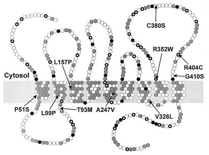
, positively charged residues; •, negatively charged residues; ○, others). Mutations identified in patients with the SLOS are indicated (arrows).

References

    1. Smith D W, Lemli L, Opitz J M. J Pediatr. 1964;64:210–217. - PubMed
    1. Tint G S, Irons M, Elias E R, Batta A K, Frieden R, Chen T S, Salen G. N Engl J Med. 1994;330:107–113. - PubMed
    1. Brown M S, Goldstein J L. Cell. 1997;89:331–340. - PubMed
    1. Moebius F F, Fitzky B U, Lee J N, Paik Y-K, Glossmann H. Proc Natl Acad Sci USA. 1998;95:1899–1902. - PMC - PubMed
    1. Budowle B, Chakraborty R, Giusti A M, Eisenberg A J, Allen R C. Am J Hum Genet. 1991;48:137–144. - PMC - PubMed

Publication types

Substances

Associated data