. 1998 Aug 4;95(16):9190-5.
doi: 10.1073/pnas.95.16.9190.

Unusual proliferation arrest and transcriptional control properties of a newly discovered E2F family member, E2F-6

Affiliations

Unusual proliferation arrest and transcriptional control properties of a newly discovered E2F family member, E2F-6

S Gaubatz et al. Proc Natl Acad Sci U S A. .

Abstract

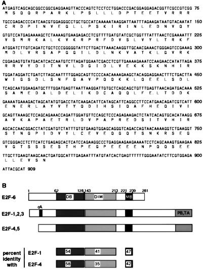
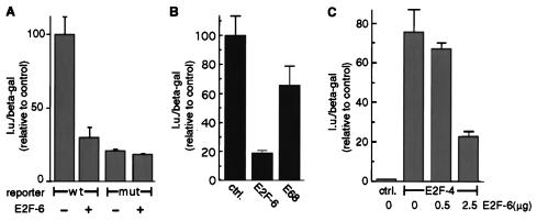
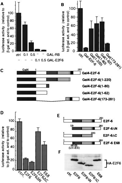
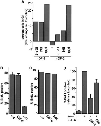
E2F transcription factors play an important role in the regulation of cell cycle progression. We report here the cloning and characterization of an additional member of this family, E2F-6. E2F-6 lacks pocket protein binding and transactivation domains, and it is a potent transcriptional repressor that contains a modular repression domain at its carboxyl terminus. Overproduction of E2F-6 had no specific effect on cell cycle progression in asynchronously growing Saos2 and NIH 3T3 cells, but it inhibited entry into S phase of NIH 3T3 cells stimulated to exit G0. Taken together, these data suggest that E2F-6 can regulate a subset of E2F-dependent genes whose products are required for entry into the cell cycle but not for normal cell cycle progression.


Figures

Figure 1
Figure 1
Human E2F-6 sequence. (A) Nucleotide sequence of a cDNA encoding human E2F-6 and the predicted amino acid sequence. The E2F-6 sequence has been submitted to the EMBL/GeneBank database (accession no. AF059292). (B) (Upper) Schematic comparison of E2F-6 with E2F-1, -2, and -3 and E2F-4 and -5. Shaded boxes indicate homologous regions. cA, cyclin A binding site; DB, DNA-binding domain; DIM, dimerization domain; MB, marked box; PB,TA, pocket protein and transactivation domain. (Lower) Homology (as expressed in percent identity in the indicated domains) between E2F-6 and E2F-1 and -4.
Figure 2
Figure 2
E2F-6 binds specifically to an E2F site. A labeled oligonucleotide corresponding to the E2F binding site in the DHFR promoter was incubated with in vitro translated myc-DP2 or HA-E2F-6 protein (lanes 1 and 2) or cotranslated myc-DP2 and HA-E2F-6 proteins (lanes 3–11). Unlabeled DHFR wild-type (wt) or mutated (mut) oligonucleotides were added in the amounts indicated. In supershift experiments, 1 μl of anti-HA antibody (HA) or anti-Myc antibody (9E10) was added. Filled arrow, position of the specific E2F-6 complex; open arrow, endogenous E2F binding activity in the reticulocyte lysate; ∗, HA supershifted band; solid circle, 9E10 supershifted band.
Figure 3
Figure 3
E2F-6 is a transcriptional repressor. (A) A luciferase reporter plasmid with three E2F consensus binding sites (wt) or mutated binding sites (mut) was cotransfected with 1 μg of E2F-6 expression plasmid or empty vector. (B) A luciferase reporter with the E2F-1 promoter was cotransfected with 1 μg of E2F-6 expression plasmid, 1 μg of E2F-6E68 (E68, DNA-binding mutant, see Fig. 4), or 1 μg of empty vector (ctrl). (C) Five micrograms of E2F-reporter plasmid was cotransfected with empty vector or 1 μg of pCDNA-E2F-4 and 0.5 or 2.5 μg of E2F-6 expression plasmid, as indicated. In all transfections, 0.2 μg of CMV-β-gal was cotransfected as internal control, and luiciferase activity was normalized to β-galactosidase activity.
Figure 4
Figure 4
E2F-6 contains a transferable repression domain. (A) Five micrograms of a luciferase reporter gene with multiple Gal4 binding sites was cotransfected with the indicated amounts of Gal4-RB or Gal4-E2F-6. (B) Five micrograms of the Gal4 luciferase reporter was cotransfected with 0.1 μg of the indicated Gal4-E2F-6 fusion protein expression vectors. (C) Schematic representation of the Gal4 fusion vectors used in A and B. (D) Five micrograms of E2F-luciferase reporter gene was cotransfected with 0.5 μg of empty vector (ctrl) or 0.5 μg of the indicated E2F-6 expression vectors. (E) Schematic representation of the E2F-6 mutants used. (F) Western blot analysis using an anti-HA antibody, showing that all E2F-6 proteins are expressed at comparable levels. In all reporter assays, 0.2 μg of CMV-β-gal was cotransfected, and luciferase activity was normalized to β-galactosidase activity.
Figure 5
Figure 5
E2F-6 does inhibit S-phase entry in starved cells but not in asynchronously growing cells. (A) Saos2 cells were cotransfected with the indicated expression vectors and CMV-GFP. Transfected cells were identified by their green fluorescence and analyzed by FACScan 2 days after transfection. Shown is the absolute change in the number of G1 cells compared with a control transfection with empty vector. The experiment was repeated four times. One typical result is shown. (B) NIH 3T3 cells were cotransfected with 1 μg of the indicated expression vectors; 0.5 μg of CMV-GFP was cotransfected. Fourteen hours after transfection, cells were labeled with BrdU for 20 hr. Cells were stained with an anti-BrdU antibody and with a rhodamine-coupled secondary antibody. Transfected cells were identified by their green fluorescence, and the number of these cells with coexisting red fluorescence (i.e., the BrdU-positive cells) was determined. The experiment was repeated several times. The mean results from one experiment performed in independent triplicates are shown. Error bars represent standard deviation. (C) NIH 3T3 cells were microinjected with 25 μg/ml E2F-6 or E2F-6E68 expression plasmid and CMV-GFP (10 μg/ml). Ten hours after microinjection, 50 μM BrdU was added for 20 hr. DNA synthesis was analyzed as described above with an anti-BrdU antibody and with a rhodamine-coupled secondary antibody. (D) E2F-6 inhibits reentry into S phase of serum-starved NIH 3T3 cells. NIH 3T3 cells were serum starved in 0.1% bovine calf serum for 48 hr and than microinjected with 25 μg/ml E2F-6 or E2F-6E68 expression plasmid. In each case CMV-GFP (10 μg/ml) was coinjected. Ten hours after injection, cells were restimulated with 5% serum, and BrdU (50 μg/ml) was added. After 17.5 hr, cells were fixed and stained with an anti-BrdU antibody as described above. The mean results from four independent experiments are shown. Values were normalized to the number of uninjected cells that had entered S phase in each experiment. In a typical experiment, 60% of serum-treated, uninjected cells had entered S phase. Error bars represent standard deviation.

References

    1. Farnham P J, Slansky J E, Kollmar R. Biochim Biophys Acta. 1993;1155:125–131. - PubMed
    1. Lam E W, La Thangue N B. Curr Opin Cell Biol. 1994;6:859–866. - PubMed
    1. Nevins J R. Science. 1992;258:424–429. - PubMed
    1. DeGregori J, Kowalk T, Nevins J R. Mol Cell Biol. 1995;15:4215–4224. - PMC - PubMed
    1. Dou Q-P, Zhao S, Levin A H, Wang J, Helin K, Pardee A B. J Biol Chem. 1994;269:1306–1313. - PubMed

Publication types

MeSH terms

Associated data

LinkOut - more resources