. 1998 Sep 15;95(19):11134-9.
doi: 10.1073/pnas.95.19.11134.

Existence of distinct tyrosylprotein sulfotransferase genes: molecular characterization of tyrosylprotein sulfotransferase-2

Affiliations

Existence of distinct tyrosylprotein sulfotransferase genes: molecular characterization of tyrosylprotein sulfotransferase-2

R Beisswanger et al. Proc Natl Acad Sci U S A. .

Abstract

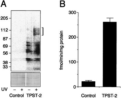
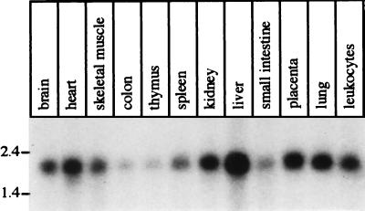
Tyrosylprotein sulfotransferase (TPST) is a 54- to 50-kDa integral membrane glycoprotein of the trans-Golgi network found in essentially all tissues investigated, catalyzing the tyrosine O-sulfation of soluble and membrane proteins passing through this compartment. Here we describe (i) an approach to identify the TPST protein, referred to as MSC (modification after substrate crosslinking) labeling, which is based on the crosslinking of a substrate peptide to TPST followed by intramolecular [35S]sulfate transfer from the cosubstrate 3'-phosphoadenosine 5'-phosphosulfate (PAPS); and (ii) the molecular characterization of a human TPST, referred to as TPST-2, whose sequence is distinct from that reported [TPST-1; Ouyang, Y.-B., Lane, W. S. & Moore, K. L. (1998) Proc. Natl. Acad. Sci. USA 95, 2896-2901] while this study was in progress. Human TPST-2 is a type II transmembrane protein of 377 aa residues that is encoded by a ubiquitously expressed 1.9-kb mRNA originating from seven exons of a gene located on chromosome 22 (22q12.1). A 304-residue segment in the luminal domain of TPST-2 shows 75% amino acid identity to the corresponding segment of TPST-1, including conservation of the residues implicated in the binding of PAPS. Expression of the TPST-2 cDNA in CHO cells resulted in an approximately 13-fold increase in both TPST protein, as determined by MSC labeling, and TPST activity. A predicted 359-residue type II transmembrane protein in Caenorhabditis elegans with 45% amino acid identity to TPST-2 in a 257-residue segment of the luminal domain points to the evolutionary conservation of the TPST protein family.


Figures

Figure 1
Figure 1
Purification of bovine TPST (A) and structure of the human TPST-2 gene and mRNA (B). (A) A mixture of TPST isolated on a preparative scale from bovine adrenal medulla by affinity chromatography on glutathione S-transferase-sulfophilin and 125I-labeled TPST purified on an analytical scale as described (17) was subjected to SDS/PAGE followed by autoradiography of the wet gel. The bracket indicates 125I-labeled TPST. The corresponding region of the gel was excised and subjected to tryptic digestion followed by HPLC separation of the peptides and microsequencing. (B) Boxes above the TPST-2 mRNA show the putative exons of the TPST-2 gene in the BAC 445C9; numbers refer to the nucleotides of the BAC 445C9 (Z95115) corresponding to the 5′ and 3′ ends of the exons. The shaded area of the TPST-2 mRNA indicates the ORF. The lines below the TPST-2 mRNA indicate some of the TPST-2 EST clones in the database. Their accession numbers are given above the lines; numbers below the lines give the 5′ and 3′ ends of the EST sequences and refer to the nucleotide sequence of the TPST-2 mRNA. The asterisks and bold accession numbers indicate the exon and EST sequences, respectively, that encode the sequence of the tryptic peptide isolated from purified bovine adrenal medullary TPST.
Figure 2
Figure 2
Comparison of the amino acid sequence of human TPST-1 (18) and TPST-2. Identical residues are shaded. Boxes indicate residues proposed (18) to be involved in PAPS binding. Bold, solid line, putative transmembrane segment of human TPST-2; triangles, exon boundaries for human TPST-2; asterisks, sequence of the tryptic peptide isolated from purified bovine adrenal medullary TPST.
Figure 3
Figure 3
Identification of TPST from bovine adrenal medulla by MSC labeling. Extracts of carbonate-treated membranes solubilized according to protocol 1 (lanes 1–4), 2 (lanes 5 and 6), or 3 (lanes 7–10) were incubated at 0°C for 30 min (lanes 5 and 6) or 1 h (lanes 1–4, 7–10) with bzbz-SgI3 (lanes 1–4) or bzbz-SgI3-HA-biotin (lanes 5–10) and [35S]PAPS without (lanes 1, 3, and 5) or with (lanes 2, 4, and 6–10) UV-irradiation, followed by further incubation either for 10 min at 0°C (0°C) or 30°C (30°C) (lanes 1–4) or for 1 h at 37°C (lanes 5 and 6) or 30°C (lanes 7–10) in the absence of UV irradiation. Samples were subjected to SDS/PAGE under reducing (lanes 1–4, 8, and 10) or nonreducing (lanes 5–7, and 9) conditions followed by transfer to poly(vinylidene difluoride) membrane and autoradiography (AR, lanes 1–8). The proteins of lanes 7 and 8 then were immunolabeled for crosslinked SgI3-HA-biotin peptide using the mAb 12CA5 against the HA epitope followed by enhanced chemiluminescence (Blot, lanes 9 and 10). Open arrows and asterisks indicate the 61- to 57-kDa TPST monomer and 122- to 114-kDa TPST dimer, respectively, bearing crosslinked [35S]sulfate-labeled SgI3 or SgI3-HA-biotin peptide.
Figure 4
Figure 4
Expression of human TPST-2 in CHO cells. CHO cells were transfected with pRC-CMV (Control) or pRC-CMV/hTPST2 (TPST-2). (A) MSC labeling. Extracts from the cell lysates (50 μg protein per lane) were incubated for 1 h at 0°C with 10 μM bzbz-SgI3-HA-biotin, 20 μM PAP, and 5 × 10⁷ cpm/ml [35S]PAPS without (−) and with (+) UV irradiation, followed by further incubation for 1 h at 30°C in the absence of UV irradiation. Samples were subjected to SDS/PAGE under nonreducing conditions followed by transfer to poly(vinylidene difluoride) membrane and autoradiography using a PhosphorImager. All four lanes of the top panel are from the same autoradiogram. The bracket indicates the 125- to 100-kDa TPST dimer bearing crosslinked [35S]sulfate-labeled SgI3-HA-biotin peptide. Note the increase in UV-independent [35S]sulfate incorporation into bands of <87 kDa in the TPST-2-transfected cells as compared with control. The two blots in the lower part of the figure show the 87- to 56-kDa region of the Ponceau S-stained poly(vinylidene difluoride) membrane, documenting the presence of equal amounts of protein. (B) Determination of TPST activity in the cell lysate. Data are the mean of two independent transfections; bars indicate the variation of the TPST activity in each cell lysate from the mean.
Figure 5
Figure 5
Northern blot analysis showing the distribution of the TPST-2 mRNA in various human tissues. Each lane contains ≈2 μg of poly(A)+ RNA.

References

    1. Huttner W B. Nature (London) 1982;299:273–276. - PubMed
    1. Niehrs C, Beisswanger R, Huttner W B. Chem Biol Interact. 1994;92:257–271. - PubMed
    1. Lee R W, Huttner W B. J Biol Chem. 1983;258:11326–11334. - PubMed
    1. Huttner W B, Niehrs C, Vannier C. Curr Biol. 1991;1:309–310. - PubMed
    1. Leyte A, van Schijndel H B, Niehrs C, Huttner W B, Verbeet M P, Mertens K, van Mourik J A. J Biol Chem. 1991;266:740–746. - PubMed

Publication types

MeSH terms

Associated data

LinkOut - more resources