. 1998 Aug 4;95(16):9572-7.
doi: 10.1073/pnas.95.16.9572.

The hereditary renal cell carcinoma 3;8 translocation fuses FHIT to a patched-related gene, TRC8

Affiliations

The hereditary renal cell carcinoma 3;8 translocation fuses FHIT to a patched-related gene, TRC8

R M Gemmill et al. Proc Natl Acad Sci U S A. .

Abstract

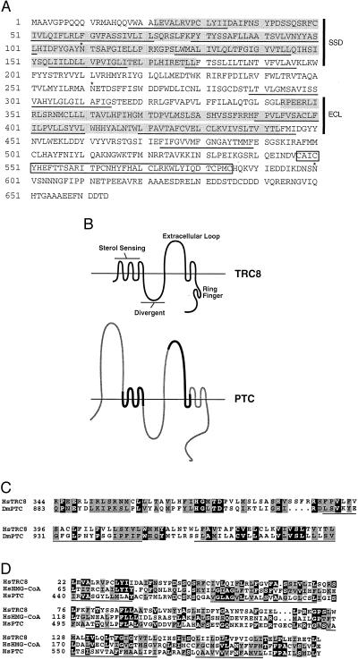
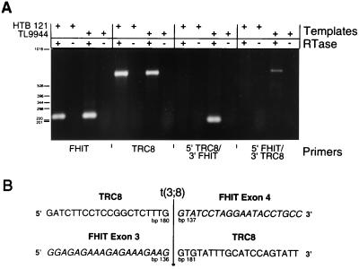
The 3;8 chromosomal translocation, t(3;8)(p14.2;q24.1), was described in a family with classical features of hereditary renal cell carcinoma. Previous studies demonstrated that the 3p14.2 breakpoint interrupts the fragile histidine triad gene (FHIT) in its 5' noncoding region. However, evidence that FHIT is causally related to renal or other malignancies is controversial. We now show that the 8q24.1 breakpoint region encodes a 664-aa multiple membrane spanning protein, TRC8, with similarity to the hereditary basal cell carcinoma/segment polarity gene, patched. This similarity involves two regions of patched, the putative sterol-sensing domain and the second extracellular loop that participates in the binding of sonic hedgehog. In the 3;8 translocation, TRC8 is fused to FHIT and is disrupted within the sterol-sensing domain. In contrast, the FHIT coding region is maintained and expressed. In a series of sporadic renal carcinomas, an acquired TRC8 mutation was identified. By analogy to patched, TRC8 might function as a signaling receptor and other pathway members, to be defined, are mutation candidates in malignant diseases involving the kidney and thyroid.


Figures

Figure 1
Figure 1
(A) Predicted amino acid sequence of TRC8. The sequence begins with the first methionine present in the isolated cDNAs. The 3;8 translocation breakpoint occurs between amino acids 60 and 61 (arrowhead). Predicted TMs are underlined, and three potential N-linked glycosylation sites are indicated by ∗. Two regions showing similarity to patched [the SSD and the extracellular loop (ECL)] are shaded. The RING-H2 motif is boxed. (B) Schematic of the predicted structure for TRC8 compared with patched (PTC). The horizontal line represents the lipid bilayer that is crossed by 10 putative TMs of TRC8 and 12 in patched. The divergent loop refers to a region of patched that is nonconserved among patched homologues (45). The diagram of patched shows regions that are similar or different from TRC8 by bold black or thin shaded lines, respectively. The N-terminal extracellular loop of patched is absent from TRC8. The predicted intracellular loop 3′ of the SSD is not conserved between either of the two known murine patched genes (Ptch1 and Ptch2) or in TRC8. (C) Amino acid sequence homology between TRC8 and Drosophila patched. The Dm Ptc sequence (GenBank accession no. M28999) was aligned with TRC8 by gapped blast. Identical amino acids are indicated by white letters on black, and similar amino acids (positive scores in a PAM250 matrix) are shaded. Two TRC8 TMs within this homology region are underlined. (D) Alignment within the putative SSD. Human sequences for HMG-CoA reductase (Swissprot accession no. P04035) and Patched (GenBank accession no. U59464) were aligned with TRC8 by gapped blast. Identities and similarities are indicated as in C; TMs within the putative SSD of TRC8 are underlined.
Figure 2
Figure 2
Analysis of TRC8 expression by Northern (A) and dot blot (B). (A) Gel resolved polyadenylated RNA (2 μg) from adult human tissues (CLONTECH) was hybridized under recommended conditions with a 1.5-kb 3′ TRC8 cDNA encompassing most of the TMs and the ring finger (bp 83–1623). A second, largely nonoverlapping probe (bp 1446–2212) yielded essentially the same pattern. The filter was exposed for 18 hr at −80°C. (B) A CLONTECH human RNA master dot blot was hybridized with the same probe as in A under recommended conditions and exposed for 15 hr. Final wash conditions were 0.1× standard saline citrate, 0.5% SDS at 55°C for 20 min. Signals were collected on a Molecular Dynamics PhosphorImager. Blank positions included B8, F5-F8, and G8. Central nervous system tissues (A1-A8 and B1-B7) included (in order) whole brain, amygdala, caudate nucleus, cerebellum, cerebral cortex, frontal lobe, hippocampus, medulla oblongata, occipital lobe, putamen, substantia nigra, temporal lobe, thalamus, sub-thalamic nucleus, and spinal cord. Musculature and digestive tissues (C1-C8) included heart, aorta, skeletal muscle, colon, bladder, uterus, prostate, and stomach. Secretory tissues (D1-D8) included testis, ovary, pancreas, pituitary, adrenal, thyroid, salivary, and mammary glands. Miscellaneous tissues (E1-E8 and F1-F4) included kidney, liver, small intestine, spleen, thymus, peripheral leukocytes, lymph node, bone marrow, appendix, lung, trachea, and placenta. Fetal tissues (G1-G7) included brain, heart, kidney, liver, spleen, thymus, and lung. All control spots (yeast and Escherichia coli RNAs, human Cot1, and total human DNAs) were blank (not shown).
Figure 3
Figure 3
(A) Localization of 5′ TRC8 sequences to chromosome 8q. Primers R-M and F-O amplify an 82-bp fragment specific for the 5′ portion of TRC8. Templates in lanes 1–11 included, respectively, AG4103 (normal human), CHO glyA (hamster), UCTP-2A3 (chromosome 3 only hybrid), 706-B6, clone17 (chromosome 8 only hybrid), TL12–8 [t(3;8) der(3) hybrid], 3;8/4–1 [t(3;8) der(8) hybrid], YAC 880A9 (chromosome 8-specific YAC spanning 3;8 breakpoint), YAC 850A6 (chromosome 3-specific YAC spanning 3;8 breakpoint), HD-7 (genomic phage clone carrying the 3;8 breakpoint region from chromosome 8), 2A7 (longest 5′RACE clone), water control. Molecular size standards are indicated in bp. (B) The same hybrid and YAC DNAs used in A were digested with EcoRI and Southern-blotted. The filter was hybridized with a 1.4-kb TRC8 cDNA fragment that derives from the 3′ end. The normal human TRC8 fragment is >15 kb, which is reduced to ≈12 kb by the translocation (arrow). The cross-hybridizing fragment in hamster DNA (lanes 2–6) is 8 kb.
Figure 4
Figure 4
(A) RT-PCR analysis of fusion product expression. RNAs isolated from the t(3;8) lymphoblastoid cell line TL9944 (36) and from a control breast carcinoma cell line (HTB121) were treated with or without RT, as indicated (+ or −) and analyzed for expression of FHIT and TRC8 by PCR. Four primers specific for 5′ and 3′ portions of each gene, F1 and R1 for FHIT and R-M and EMR for TRC8, were used in combination to detect both wild-type and putative chimeric transcripts. The FHIT primer pair generated a product of the expected size (231 bp) as did the TRC8 primer pair (651 bp). Reciprocal chimeric products were amplified by using R-M plus R1 for 5′ TRC8/3′ FHIT and F1 plus EMR for 5′ FHIT/3′ TRC8. Predicted sizes of the chimeric products are 188 and 694 bp, respectively. (B) Sequences of 3;8 chimeric transcripts. The RT-PCR-amplified cDNAs in lanes 11 and 15, corresponding to the reciprocal chimeric transcripts, were purified and sequenced on both strands. Forty bp surrounding the boundary between FHIT exons 3 and 4 are shown with FHIT sequences italicized. The precise position of the fusion on both TRC8 and FHIT transcripts is indicated.
Figure 5
Figure 5
(A) Detection of a tumor-specific somatic mutation by SSCP and heteroduplex analysis. DNA samples were PCR-amplified by using primers flanking the first coding exon of TRC8. The products were denatured, separated on a nondenaturing MDE gel and detected by silver staining. Samples included matched tumor and normal DNAs from patients 1 and 7 (lanes 5–8, respectively) and an unrelated normal control (AG4103, lane 9). A separate SSCP gel was used to isolate four individual SSCP bands from RCC no. 1 (lane 5, marked by an arrow or arrowheads). The excised bands A-D corresponded to the indicated bands in lane 5 from top to bottom. These last templates were reamplified and analyzed by SSCP to determine whether they contained mutant or wild-type sequences. Comparison to lane 5 suggested that bands A and C contained primarily mutant DNA, band B was a mixture of mutant and wild-type, and band D was wild-type only. Results for SSCP (Upper) and heteroduplex (Lower) are shown. (B) RCC no. 1 contains a 12-bp duplication in the 5′ untranslated region. Purified PCR products shown in A (lanes 1–4) were sequenced. The mutation consisted of a 12-bp direct duplication (underlined) at bp position −73, which was present in the tumor sample but not in the corresponding normal DNA.

References

    1. Cohen A J, Li F P, Berg S, Marchetto D J, Tsai S, Jacobs S C, Brown R S. N Engl J Med. 1979;301:592–595. - PubMed
    1. Li F P, Decker H J, Zbar B, Stanton V P, Jr, Kovacs G, Seizinger B R, Aburatani H, Sandberg A A, Berg S, Hosoe S. Ann Intern Med. 1993;118:106–111. - PubMed
    1. Latif F, Tory K, Gnarra J, Yao M, Duh F M, Orcutt M L, Stackhouse T, Kuzmin I, Modi W, Geil L, et al. Science. 1993;260:1317–1320. - PubMed
    1. van den Berg A, Buys C H. Genes Chromosomes Cancer. 1997;19:59–76. - PubMed
    1. Ohta M, Inoue H, Cotticelli M G, Kastury K, Baffa R, Palazzo J, Siprashvili Z, Mori M, McCue P, Druck T, et al. Cell. 1996;84:587–597. - PubMed

Publication types

MeSH terms

Associated data