. 1999 Dec;67(12):6533-42.
doi: 10.1128/IAI.67.12.6533-6542.1999.

The pspC gene of Streptococcus pneumoniae encodes a polymorphic protein, PspC, which elicits cross-reactive antibodies to PspA and provides immunity to pneumococcal bacteremia

Affiliations

The pspC gene of Streptococcus pneumoniae encodes a polymorphic protein, PspC, which elicits cross-reactive antibodies to PspA and provides immunity to pneumococcal bacteremia

A Brooks-Walter et al. Infect Immun. 1999 Dec.

Abstract

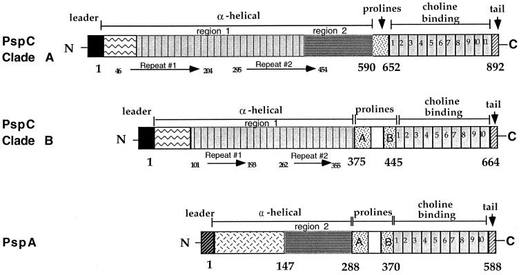
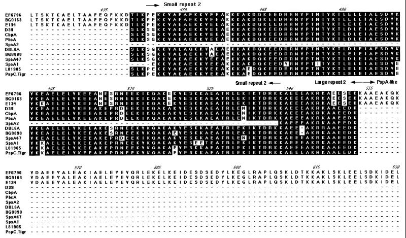
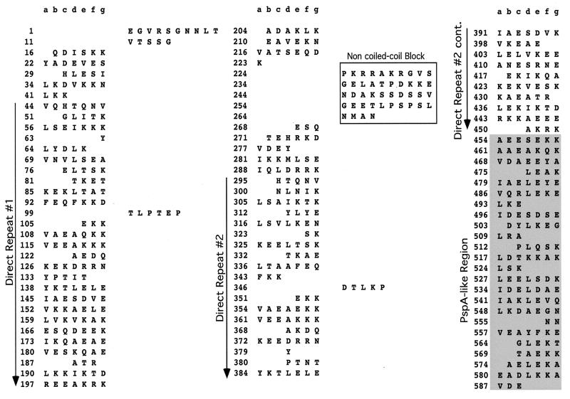
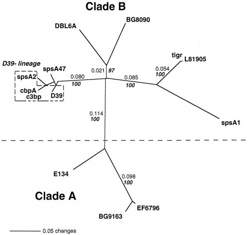
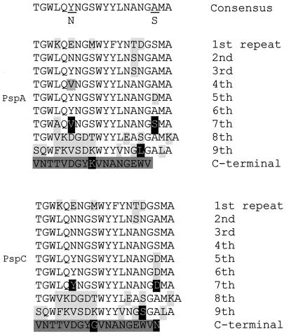
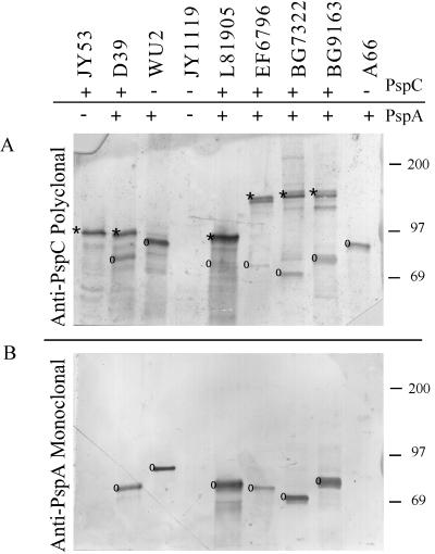
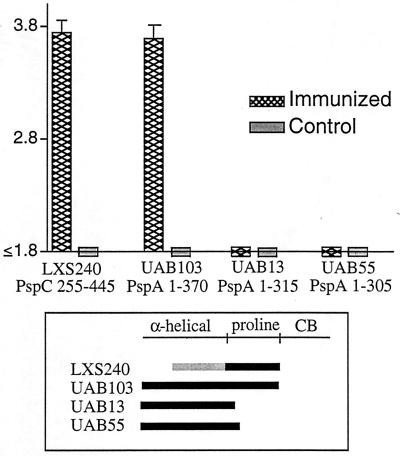
PspC is one of three designations for a pneumococcal surface protein whose gene is present in approximately 75% of all Streptococcus pneumoniae strains. Under the name SpsA, the protein has been shown to bind secretory immunoglobulin A (S. Hammerschmidt, S. R. Talay, P. Brandtzaeg, and G. S. Chhatwal, Mol. Microbiol. 25:1113-1124, 1997). Under the name CbpA, the protein has been shown to interact with human epithelial and endothelial cells (C. Rosenow et al., Mol. Microbiol. 25:819-829, 1997). The gene is paralogous to the pspA gene in S. pneumoniae and was thus called pspC (A. Brooks-Walter, R. C. Tart, D. E. Briles, and S. K. Hollingshead, Abstracts of the 97th General Meeting of the American Society for Microbiology 1997). Sequence comparisons of five published and seven new alleles reveal that this gene has a mosaic structure, and modular domains have contributed to gene diversity during evolution. Two major clades exist: clade A alleles are larger and contain an extra module that is shared with many pspA alleles; clade B alleles are smaller and lack this pspA-like domain. All alleles have a proline-rich domain and a choline-binding repeat domain that show 0% divergence from similar domains in the PspA protein. Immunization of a rabbit with a recombinant clade B PspC molecule produced antiserum that cross-reacted with both PspC and PspA from 15 pneumococcal isolates. The cross-reactive antibodies afforded cross-protection in a mouse model system. Mice immunized with PspC were protected against challenge with a strain that expressed PspA but not PspC. The PspA- and PspC-cross-reactive antibodies were directed to the proline-rich domain present in both molecules.


Figures

FIG. 1
FIG. 1
Cartoon of the PspC clades compared to a representative PspA molecule. Long arrows represent the direct repeats found within the α helix. The hypervariable region is indicated by the box containing zigzag lines. The region showing homology to the α helix is indicated by the box containing horizontal lines.
FIG. 2
FIG. 2
Alignment of PspC. The amino acid sequences which included the α-helical region and the proline-rich region of PspC were aligned with Mac Vector 6.5. The direct repeats within the α helix, the non-coiled-coil block, and the proline-rich region are indicated with arrows. Conserved regions are shaded, and gaps are shown with a dash. Entries are named for the strain from which the gene was cloned, with the exception of GenBank entries: SpsA1 (Y10818) from strain ATCC 33400 (serotype 1), SpsA2 (AJ002054) from strain ATCC 11733 (serotype 2), SpsA47 (AJ002055) from strain NCTC10319 (serotype 47), CbpA (AF019904) from strain LM91 (serotype 2), PbcA (a C3-binding protein) (AF067128), and TIGR sequence for a serotype 4 clinical isolate (30a). The capsular serotypes of the other strains are as follows: EF6796, 6A; BG8090, 19; L81905, 4; DBL6A, 6A; BG9163, 6B; D39, 2; and E134, 23.
FIG. 2
FIG. 2
Alignment of PspC. The amino acid sequences which included the α-helical region and the proline-rich region of PspC were aligned with Mac Vector 6.5. The direct repeats within the α helix, the non-coiled-coil block, and the proline-rich region are indicated with arrows. Conserved regions are shaded, and gaps are shown with a dash. Entries are named for the strain from which the gene was cloned, with the exception of GenBank entries: SpsA1 (Y10818) from strain ATCC 33400 (serotype 1), SpsA2 (AJ002054) from strain ATCC 11733 (serotype 2), SpsA47 (AJ002055) from strain NCTC10319 (serotype 47), CbpA (AF019904) from strain LM91 (serotype 2), PbcA (a C3-binding protein) (AF067128), and TIGR sequence for a serotype 4 clinical isolate (30a). The capsular serotypes of the other strains are as follows: EF6796, 6A; BG8090, 19; L81905, 4; DBL6A, 6A; BG9163, 6B; D39, 2; and E134, 23.
FIG. 3
FIG. 3
Coiled-coil motif of the α helix of EF6796 PspC. Amino acids that are not in the coiled-coil motif are shifted to the right column. The direct repeats of the α helix and the non-coiled-coil block are indicated. The region with homology to PspA is shaded. This is the output from the Matcher program (10, 24a).
FIG. 4
FIG. 4
Tree of the PspC proteins from this study and related proteins SpsA and CbpA from GenBank. PspC proteins were truncated after the proline-rich region (Fig. 1) before being aligned with the Clustal W algorithm and the Blosum30 amino-acid-scoring matrix in Mac Vector. The tree is an unrooted phylogram generated by the neighbor-joining method with mean character distances in the program PAUP 4.0B. Nonitalic numbers on the tree indicate distances along the branch lengths as calculated by PAUP. Italic bold numbers indicate the percentage of time that the branches were joined together under bootstrap analysis (1,000 replicates were performed). Clade A and clade B are monophyletic groups which were separated by a distance of greater than 0.1 and which clustered together 100% of the time. Clade A PspC proteins share a 120-aa domain with many PspA proteins (Fig. 2). Clade B PspC proteins lack the 120-aa domain, but the other PspC, SpsA, or CbpA proteins share the proline-rich domain with the PspA proteins. The boxed D39 lineage indicates different sequences originating from strains that are laboratory descendents of strain D39. The entries used were the same as those described in the legend to Fig. 2.
FIG. 5
FIG. 5
Consensus sequences for choline-binding regions of PspC and PspA. The repeat regions of eight proteins, which included three PspA and five PspC molecules, were aligned with the Clustal W algorithm. The alignment was adjusted to maintain divergent repeats together to account for the various sizes of the choline-binding domains. The consensus for repeat number 1 through repeat number 9 of the PspA proteins is in the upper half of figure and that of the PspC proteins is in the bottom half of the figure. The consensus for all nine repeats (1–9) representing at least 60% of the individual amino acids in each position is shown at the top of the diagram. The amino acids that diverge from the consensus sequence are indicated in light gray, and the C-terminal tail is indicated in dark gray. Five amino acids that vary in a gene-specific manner are indicated in white letters on a black background.
FIG. 6
FIG. 6
Western immunoblot of pneumococcal lysates. Panel A was developed with anti-PspC polyclonal serum, and panel B was developed with anti-PspA MAb Xi126. An asterisk indicates PspC proteins, and a circle indicates PspA proteins. Molecular mass markers (in kilodaltons) are indicated on the right. Cross-reaction of the polyclonal serum to PspC was observed with all strains tested, but the polyclonal serum reacted weakly to the PspA molecule from L81905.
FIG. 7
FIG. 7
Antibody response patterns in cross-protection. The antibody responses to recombinant PspC and PspA in sera of mice immunized with PspC were measured by an ELISA, and log reciprocal titers were determined. Each bar on the graph represents the mean of the log reciprocal titer and the upper boundary of the standard error for sera from five mice. The limit of detection for the log reciprocal antibody titers in these assays was 1.8. CB, choline-binding region.

References

    1. Ausubel F M, Brent R, Kingston R E, Moore D D, Seidman J G, Smith J A, Struhl K, editors. Current protocols in molecular biology. Vol. 1. New York, N.Y: John Wiley & Sons, Inc.; 1987.
    1. Banas J A, Russell R R B, Ferretti J J. Sequence analysis of the gene for the glucan-binding protein of Streptococcus mutansIngbritt. Infect Immun. 1990;58:667–673. - PMC - PubMed
    1. Barroso L A, Wang S-Z, Phelps C J, Johnson J L, Wilkins T D. Nucleotide sequence of Clostridium difficiletoxin B gene. Nucleic Acids Res. 1990;18:4004. - PMC - PubMed
    1. Breise T, Hackenbeck R. Interactions of pneumococcal amidase with lipoteichoic acid and choline. Eur J Biochem. 1985;146:417–427. - PubMed
    1. Briles D E, Crain M J, Gray B M, Forman C, Yother J. A strong association between capsular type and mouse virulence among human isolates of Streptococcus pneumoniae. Infect Immun. 1992;60:111–116. - PMC - PubMed

Publication types

MeSH terms

LinkOut - more resources