. 1998 Nov;72(11):8586-96.
doi: 10.1128/JVI.72.11.8586-8596.1998.

Protection against fatal Sindbis virus encephalitis by beclin, a novel Bcl-2-interacting protein

Affiliations

Protection against fatal Sindbis virus encephalitis by beclin, a novel Bcl-2-interacting protein

X H Liang et al. J Virol. 1998 Nov.

Abstract

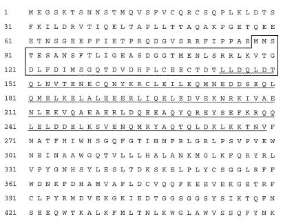
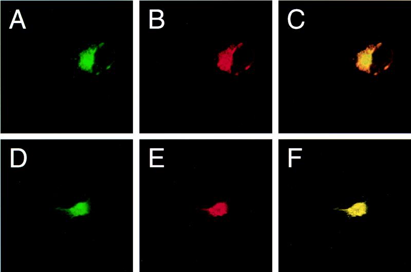
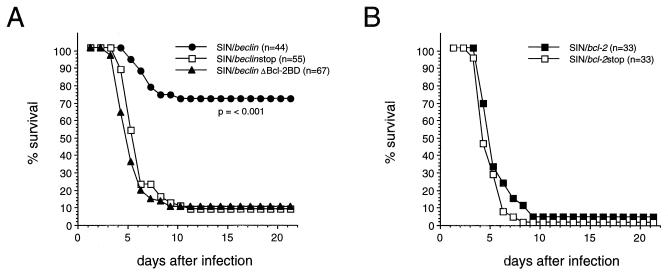
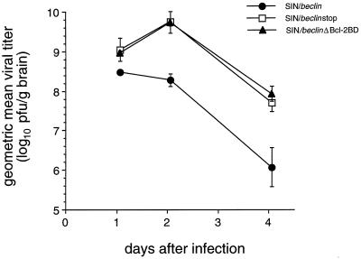
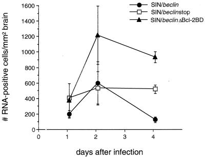
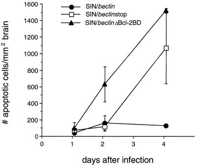
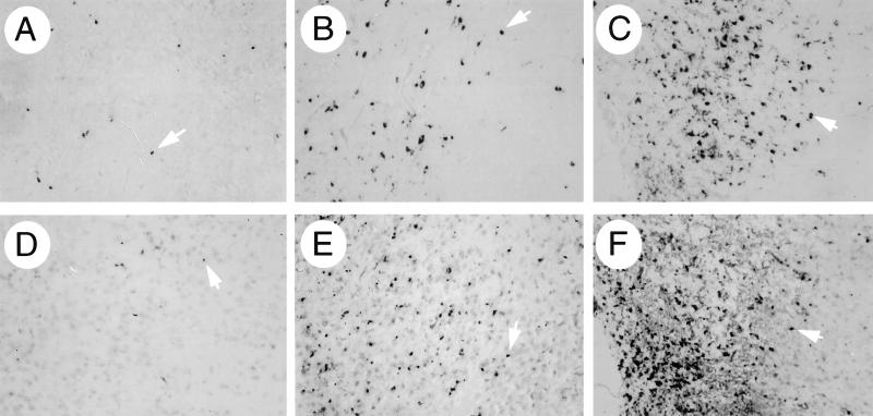
bcl-2, the prototypic cellular antiapoptotic gene, decreases Sindbis virus replication and Sindbis virus-induced apoptosis in mouse brains, resulting in protection against lethal encephalitis. To investigate potential mechanisms by which Bcl-2 protects against central nervous system Sindbis virus infection, we performed a yeast two-hybrid screen to identify Bcl-2-interacting gene products in an adult mouse brain library. We identified a novel 60-kDa coiled-coil protein, Beclin, which we confirmed interacts with Bcl-2 in mammalian cells, using fluorescence resonance energy transfer microscopy. To examine the role of Beclin in Sindbis virus pathogenesis, we constructed recombinant Sindbis virus chimeras that express full-length human Beclin (SIN/beclin), Beclin lacking the putative Bcl-2-binding domain (SIN/beclinDeltaBcl-2BD), or Beclin containing a premature stop codon near the 5' terminus (SIN/beclinstop). The survival of mice infected with SIN/beclin was significantly higher (71%) than the survival of mice infected with SIN/beclinDeltaBcl-2BD (9%) or SIN/beclinstop (7%) (P < 0.001). The brains of mice infected with SIN/beclin had fewer Sindbis virus RNA-positive cells, fewer apoptotic cells, and lower viral titers than the brains of mice infected with SIN/beclinDeltaBcl-2BD or SIN/beclinstop. These findings demonstrate that Beclin is a novel Bcl-2-interacting cellular protein that may play a role in antiviral host defense.


Figures

FIG. 1
FIG. 1
Deduced amino acid sequence of human Beclin. The boxed area represents the Bcl-2-binding domain of human Beclin (Table 1), and the underlined area corresponds to the region that is predicted to have a coiled-coil conformation.
FIG. 2
FIG. 2
Beclin mRNA and protein expression. (A) Northern blot analysis of beclin mRNA expression in human and mouse tissues. sm., small; m., muscle. (B) Immunoperoxidase staining of an adult human hippocampus section with a polyclonal antibody against a human Beclin peptide. The arrow marks a Beclin-positive neuron. Magnification, ×450.
FIG. 2
FIG. 2
Beclin mRNA and protein expression. (A) Northern blot analysis of beclin mRNA expression in human and mouse tissues. sm., small; m., muscle. (B) Immunoperoxidase staining of an adult human hippocampus section with a polyclonal antibody against a human Beclin peptide. The arrow marks a Beclin-positive neuron. Magnification, ×450.
FIG. 3
FIG. 3
Expression of flag-Beclin protein constructs by the virus vectors SIN/beclin, SIN/beclinstop, and SIN/beclinΔBcl-2BD. (A and B) Western blot analyses of virus-infected BHK cell lysates with either anti-flag epitope antibody M2 (A) or polyclonal rabbit anti-Beclin antiserum (B). Lane 1, SIN/beclin; lane 2, SIN/beclinstop; lane 3, SIN/beclinΔBcl-2BD; lane 4, empty Sindbis virus vector. (C to E) Immunoperoxidase staining using anti-flag epitope antibody M2 of mouse brains 2 days after infection with SIN/beclin (C), SIN/beclinstop (D), or SIN/beclinΔBcl-2BD (E). Magnification, ×111.
FIG. 4
FIG. 4
Confocal laser scanning microscopy of COS7 cells expressing Bcl-2 and Beclin. (A and B) Cell cotransfected with pSG5/bcl-2 and pSG5/flag-beclin and stained with anti-flag epitope (A) and anti-human Bcl-2 (B) antibodies. (D and E) Cell cotransfected with pSG5/bcl-2 and pSG5/flag-beclinDBcl-2BD and stained with anti-flag epitope (D) and anti-human Bcl-2 (E) antibodies. (C and F) Computerized overlays of panels A and B (C) and panels D and E (F); yellow color corresponds to overlap of FITC and rhodamine staining. Confocal slides were 1 μm thick.
FIG. 5
FIG. 5
Effects of Beclin and Bcl-2 on the survival of mice infected with Sindbis virus strain TE12. (A) Survival curve of mice infected with SIN/beclin, SIN/beclinstop, and SIN/beclinΔBcl-2BD. Data represent combined survival probabilities for four to six independent litters. (B) Survival curve of mice infected with SIN/bcl-2 and SIN/bcl-2stop. Data represent combined survival probabilities for three independent litters.
FIG. 6
FIG. 6
Viral growth of SIN/beclin, SIN/beclinstop, and SIN/beclinΔBcl-2BD in mouse brain. Each data point represents geometric mean viral titer ± SEM of three to six mouse brains.
FIG. 7
FIG. 7
Viral RNA-positive cells in mouse brains infected with SIN/beclin, SIN/beclinstop, and SIN/beclinΔBcl-2BD. Each data point represents the mean ± SEM number of Sindbis virus message-sense RNA-positive cells per square millimeter of brain for three to six mouse brains.
FIG. 8
FIG. 8
Apoptotic nuclei in mouse brains infected with SIN/beclin, SIN/beclinstop, and SIN/beclinΔBcl-2BD. Each data point represents the mean ± SEM number of ISEL-positive nuclei per square millimeter of brain for three to six mouse brains.
FIG. 9
FIG. 9
Representative photomicrographs of fields used for computerized quantitative image analysis of viral RNA (A to C)- and ISEL (D to F)-positive cells in mouse brains infected with SIN/beclin (A and D), SIN/beclinstop (B and E), and SIN/beclinΔBcl-2BD (C and F) in Fig. 7 and 8, respectively. All photomicrographs are from the colliculus region of brain sections 4 days after infection and correspond to the images observed at a magnification of ×10. Arrowheads denote representative cells that were scored as positive by the Image-ProPlus software program.

References

    1. Allsopp T E, Scallan M F, Williams A, Fazakerley J K. Virus infection induces neuronal apoptosis: a comparison with trophic factor withdrawal. Cell Death Differ. 1998;5:50–59. - PubMed
    1. Altshul S F, Gish W, Miller W, Meyers E W, Lipman D J. Basic local alignment tool. J Mol Biol. 1990;215:403–410. - PubMed
    1. Antoni B A, Sabbatini P A, Rabson A B, White E. Inhibition of apoptosis in human immunodeficiency virus-infected cells enhances virus production and facilitates persistent infection. J Virol. 1995;69:2384–2392. - PMC - PubMed
    1. Boise L H, Gonzalez-Garcia M, Postema C E, Ding L, Lindsten T, Turka L A, Mao X, Nunez G, Thompson C B. Bcl-xL, a bcl-2 related gene that functions as a dominant regulator of apoptotic cell death. Cell. 1993;74:597–608. - PubMed
    1. Cheng E H Y, Levine B, Boise L H, Thompson C B, Hardwick J M. Bax-independent inhibition of apoptosis by Bcl-xL. Nature. 1996;379:554–556. - PubMed

Publication types

MeSH terms

Associated data