. 1998 Nov 16;188(10):1841-8.
doi: 10.1084/jem.188.10.1841.

Mouse CD94/NKG2A is a natural killer cell receptor for the nonclassical major histocompatibility complex (MHC) class I molecule Qa-1(b)

Affiliations

Mouse CD94/NKG2A is a natural killer cell receptor for the nonclassical major histocompatibility complex (MHC) class I molecule Qa-1(b)

R E Vance et al. J Exp Med. .

Abstract

Natural killer (NK) cells preferentially lyse targets that express reduced levels of major histocompatibility complex (MHC) class I proteins. To date, the only known mouse NK receptors for MHC class I belong to the Ly49 family of C-type lectin homodimers. Here, we report the cloning of mouse NKG2A, and demonstrate it forms an additional and distinct class I receptor, a CD94/NKG2A heterodimer. Using soluble tetramers of the nonclassical class I molecule Qa-1(b), we provide direct evidence that CD94/NKG2A recognizes Qa-1(b). We further demonstrate that NK recognition of Qa-1(b) results in the inhibition of target cell lysis. Inhibition appears to depend on the presence of Qdm, a Qa-1(b)-binding peptide derived from the signal sequences of some classical class I molecules. Mouse NKG2A maps adjacent to CD94 in the heart of the NK complex on mouse chromosome six, one of a small cluster of NKG2-like genes. Our findings suggest that mouse NK cells, like their human counterparts, use multiple mechanisms to survey class I expression on target cells.


Figures

Figure 1
Figure 1
Comparison of mouse (m), rat (r), and human (h) NKG2A protein sequences, as deduced from the cDNA sequences, using the Clustal W algorithm (v 1.6; reference 52). Identical residues are joined by a vertical line and similar residues are joined by a colon. The exon structure of the human NKG2A gene is shown, as determined previously (53). Potential or actual ITIM are shaded, as are the predicted transmembrane domains and potential N-linked glycosylation sites (*). The mouse NKG2A nucleotide sequence is available from GenBank under accession number AF095447.
Figure 2
Figure 2
The NKG2 multi-gene family genomic structure. (A) Tail genomic DNA was isolated from the indicated strains of inbred mice, digested with EcoRI or BamHI, and Southern blotted with a probe derived from predicted exons 5 and 6 of mouse NKG2A. The migration of standard kilobase markers is shown. (B) Hybridization of NK complex probes to BACs. Clone numbers (Genome Systems B6 BAC library) and restriction enzymes used in the digest are shown above each lane. NotI was added to the EcoRV and EcoRI digests to ensure complete release of the insert; HindIII is sufficient to release the insert. 95R is an NK complex marker previously derived from the right end of YAC 95E6 (24). See Materials and Methods for the details of other probes. The NKG2A 5′ probe might be weakly cross-reactive with another NKG2 family member, as evidenced by its weak hybridization to a 2-kb EcoRV fragment in BACs 189c16 and 89b04. (C) Proposed structure of the mouse CD94/ NKG2 complex, located on chromosome six between the Nkrp1 and Cd69 genes (centromerically) and the Ly49 cluster (telomerically). BACs were ordered on the basis of the hybridization data in B. The number and order of NKG2C/E genes remains uncertain, and awaits the development of more specific probes.
Figure 3
Figure 3
CD94/NKG2A is a receptor for Qa-1b. Expression constructs encoding CD94, NKG2A, or HA epitope-tagged versions of these proteins, were transiently transfected individually or together into COS7 cells as indicated. After 48 h, cells were lifted with PBS and 0.02% EDTA, stained either with anti-HA or Qa-1b tetramer, and analyzed by flow cytometry using forward and side scatter to exclude dead cells. The percentage of cells within the indicated gate is shown for each sample.
Figure 4
Figure 4
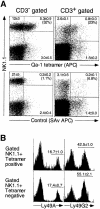
The Qa-1b tetramer predominantly recognizes a subset of NK cells. (A) B6 splenocytes were enriched for T and NK cells by passage over nylon wool and stained with the indicated monoclonal antibodies and with streptavidin-allophycocyanin (SAv-APC) complexed Qa-1b tetramer (top). As a control for the specificity of the tetramer, cells were also stained with streptavidin-APC alone (bottom). Left hand panels are gated on CD3 negative cells, whereas right hand panels are gated on CD3 positive cells. The percentages of gated cells in each quadrant are indicated, and are the average of 3 mice ± SD. The percentage of APC+ cells among NK1.1+ cells is given in parentheses. (B) The receptor for Qa-1b on NK cells only partially overlaps in its expression with other class I–specific receptors on NK cells. NK1.1+/tetramer positive or negative cells were electronically gated and analyzed for their binding of FITC-conjugated monoclonal antibodies to Ly49 family members. Again, the percentage of cells within the indicated gate is given ± SD (n = 3 mice).
Figure 5
Figure 5
(A) Inhibition of NK cells by recognition of Qa-1/Qdm. Pure IL-2 cultured NK cells (CD3NK1.1+) cells were sorted for the expression (Qa-1tet positive) or the lack of expression (Qa-1tet negative) of Qa-1b receptor and assayed for cytotoxicity against 51Cr labeled T2 cells stably transfected with Qa-1b (T2-37, top) or IAb (T2-BB, bottom). Targets were incubated overnight in media at 26°C with the addition of excess (30 μM) peptide or an equal volume of PBS. Ova (SIINFEKL) is a Kb binding peptide; Qdm (AMAPRTLLL) is a Qa-1b binding peptide. The kill was performed in the presence of the peptide/PBS at the indicated E:T ratios. (B) Treatment with Qdm peptide does not detectably enhance surface expression of Qa-1b in T2-Qa-1b transfectants (T2-37 cells). T2-37 cells were incubated overnight at 26°C in the presence of 30 μM peptide or PBS. To control for the 37°C incubation required to label the targets with 51Cr (see A), the T2-37 cells were then incubated at 37°C for 1 h in the absence of   51Cr but in the presence of appropriate peptide. Cells were then stained with normal rabbit serum (NRS, dotted histograms) or a specific rabbit anti-Qa-1b polyclonal antiserum (filled histograms) at 4°C in the presence of sodium azide (0.02%), followed by staining with FITC-conjugated swine anti–rabbit F(ab′)2. Numbers indicate the mean fluorescence intensity of the population.

References

    1. Ehrlich R. Modulation of antigen processing and presentation by persistent virus infections and in tumors. Hum Immunol. 1997;54:104–116. - PubMed
    1. Ploegh HL. Viral strategies of immune evasion. Science. 1998;280:248–253. - PubMed
    1. Ljunggren HG, Karre K. In search of the ‘missing self': MHC molecules and NK cell recognition. Immunol Today. 1990;11:237–244. - PubMed
    1. Yokoyama WM. Hybrid resistance and the Ly-49 family of natural killer cell receptors. J Exp Med. 1995;182:273–277. - PMC - PubMed
    1. Vance RE, Raulet DH. Toward a quantitative analysis of the repertoire of class I MHC-specific inhibitory receptors on natural killer cells. Curr Top Microbiol Immunol. 1998;230:135–160. - PubMed

Publication types

Associated data