. 1998 Dec 8;95(25):14717-22.
doi: 10.1073/pnas.95.25.14717.

WISP genes are members of the connective tissue growth factor family that are up-regulated in wnt-1-transformed cells and aberrantly expressed in human colon tumors

Affiliations

WISP genes are members of the connective tissue growth factor family that are up-regulated in wnt-1-transformed cells and aberrantly expressed in human colon tumors

D Pennica et al. Proc Natl Acad Sci U S A. .

Abstract

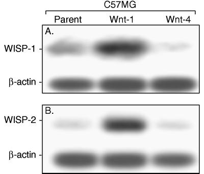
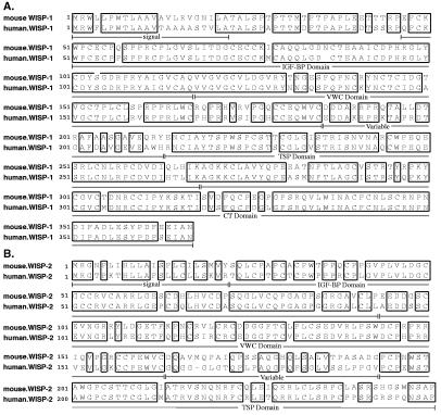
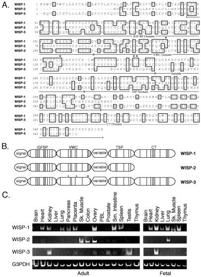
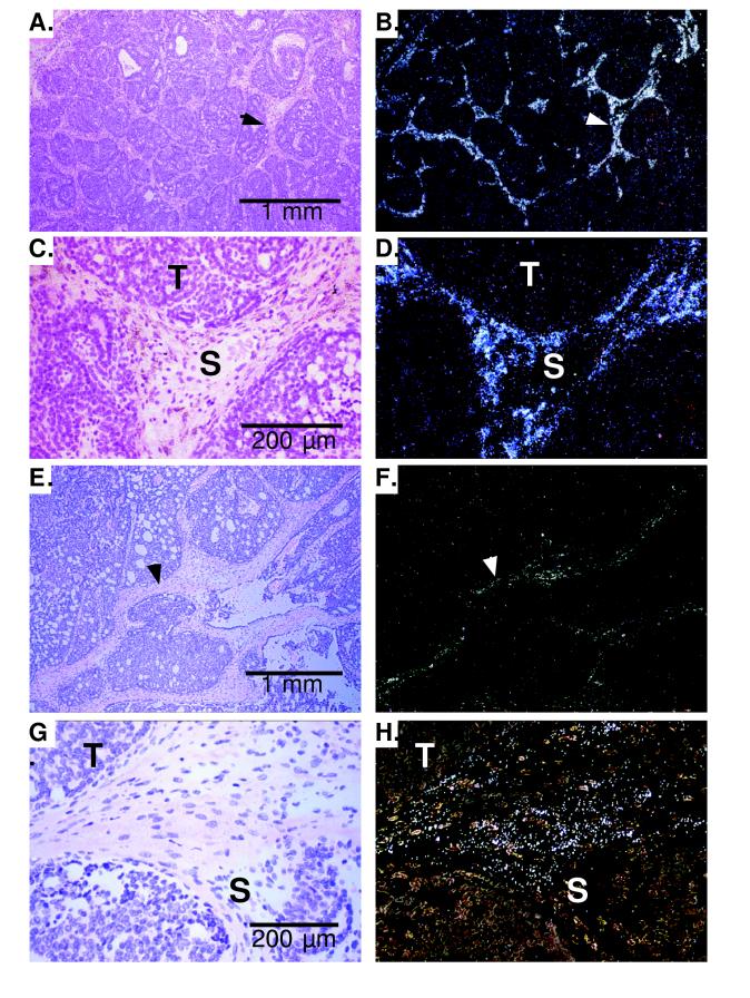
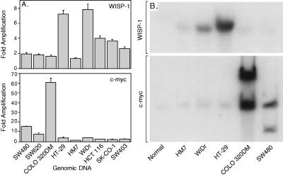
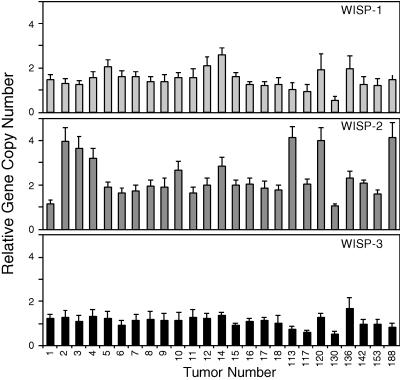
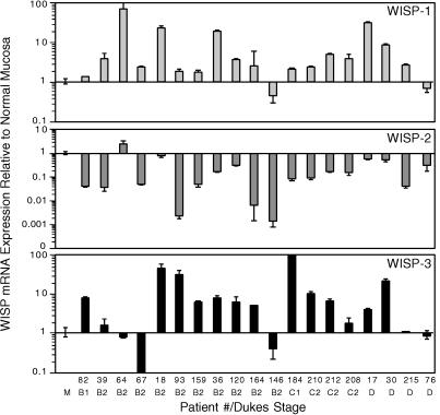
Wnt family members are critical to many developmental processes, and components of the Wnt signaling pathway have been linked to tumorigenesis in familial and sporadic colon carcinomas. Here we report the identification of two genes, WISP-1 and WISP-2, that are up-regulated in the mouse mammary epithelial cell line C57MG transformed by Wnt-1, but not by Wnt-4. Together with a third related gene, WISP-3, these proteins define a subfamily of the connective tissue growth factor family. Two distinct systems demonstrated WISP induction to be associated with the expression of Wnt-1. These included (i) C57MG cells infected with a Wnt-1 retroviral vector or expressing Wnt-1 under the control of a tetracyline repressible promoter, and (ii) Wnt-1 transgenic mice. The WISP-1 gene was localized to human chromosome 8q24.1-8q24.3. WISP-1 genomic DNA was amplified in colon cancer cell lines and in human colon tumors and its RNA overexpressed (2- to >30-fold) in 84% of the tumors examined compared with patient-matched normal mucosa. WISP-3 mapped to chromosome 6q22-6q23 and also was overexpressed (4- to >40-fold) in 63% of the colon tumors analyzed. In contrast, WISP-2 mapped to human chromosome 20q12-20q13 and its DNA was amplified, but RNA expression was reduced (2- to >30-fold) in 79% of the tumors. These results suggest that the WISP genes may be downstream of Wnt-1 signaling and that aberrant levels of WISP expression in colon cancer may play a role in colon tumorigenesis.


Figures

Figure 1
Figure 1
WISP-1 and WISP-2 are induced by Wnt-1, but not Wnt-4, expression in C57MG cells. Northern analysis of WISP-1 (A) and WISP-2 (B) expression in C57MG, C57MG/Wnt-1, and C57MG/Wnt-4 cells. Poly(A)+ RNA (2 μg) was subjected to Northern blot analysis and hybridized with a 70-bp mouse WISP-1-specific probe (amino acids 278–300) or a 190-bp WISP-2-specific probe (nucleotides 1438–1627) in the 3′ untranslated region. Blots were rehybridized with human β-actin probe.
Figure 2
Figure 2
Encoded amino acid sequence alignment of mouse and human WISP-1 (A) and mouse and human WISP-2 (B). The potential signal sequence, insulin-like growth factor-binding protein (IGF-BP), VWC, thrombospondin (TSP), and C-terminal (CT) domains are underlined.
Figure 3
Figure 3
(A) Encoded amino acid sequence alignment of human WISPs. The cysteine residues of WISP-1 and WISP-2 that are not present in WISP-3 are indicated with a dot. (B) Schematic representation of the WISP proteins showing the domain structure and cysteine residues (vertical lines). The four cysteine residues in the VWC domain that are absent in WISP-3 are indicated with a dot. (C) Expression of WISP mRNA in human tissues. PCR was performed on human multiple-tissue cDNA panels (CLONTECH) from the indicated adult and fetal tissues.
Figure 4
Figure 4
(A, C, E, and G) Representative hematoxylin/eosin-stained images from breast tumors in Wnt-1 transgenic mice. The corresponding dark-field images showing WISP-1 expression are shown in B and D. The tumor is a moderately well-differentiated adenocarcinoma showing evidence of adenoid cystic change. At low power (A and B), expression of WISP-1 is seen in the delicate branching fibrovascular tumor stroma (arrowhead). At higher magnification, expression is seen in the stromal(s) fibroblasts (C and D), and tumor cells are negative. Focal expression of WISP-1, however, was observed in tumor cells in some areas. Images of WISP-2 expression are shown in EH. At low power (E and F), expression of WISP-2 is seen in cells lying within the fibrovascular tumor stroma. At higher magnification, these cells appeared to be adjacent to capillary vessels whereas tumor cells are negative (G and H).
Figure 5
Figure 5
Amplification of WISP-1 genomic DNA in colon cancer cell lines. (A) Amplification in cell line DNA was determined by quantitative PCR. (B) Southern blots containing genomic DNA (10 μg) digested with EcoRI (WISP-1) or XbaI (c-myc) were hybridized with a 100-bp human WISP-1 probe (amino acids 186–219) or a human c-myc probe (located at bp 1901–2000). The WISP and myc genes are detected in normal human genomic DNA after a longer film exposure.
Figure 6
Figure 6
Genomic amplification of WISP genes in human colon tumors. The relative gene copy number of the WISP genes in 25 adenocarcinomas was assayed by quantitative PCR, by comparing DNA from primary human tumors with pooled DNA from 10 healthy donors. The data are means ± SEM from one experiment done in triplicate. The experiment was repeated at least three times.
Figure 7
Figure 7
WISP RNA expression in primary human colon tumors relative to expression in normal mucosa from the same patient. Expression of WISP mRNA in 19 adenocarcinomas was assayed by quantitative PCR. The Dukes stage of the tumor is listed under the sample number. The data are means ± SEM from one experiment done in triplicate. The experiment was repeated at least twice.

References

    1. Cadigan K M, Nusse R. Genes Dev. 1997;11:3286–3305. - PubMed
    1. Dale T C. Biochem J. 1998;329:209–223. - PMC - PubMed
    1. Nusse R, Varmus H E. Cell. 1982;31:99–109. - PubMed
    1. van Ooyen A, Nusse R. Cell. 1984;39:233–240. - PubMed
    1. Tsukamoto A S, Grosschedl R, Guzman R C, Parslow T, Varmus H E. Cell. 1988;55:619–625. - PubMed

MeSH terms