. 1999 May 11;96(10):5616-21.
doi: 10.1073/pnas.96.10.5616.

An imprinted, mammalian bicistronic transcript encodes two independent proteins

Affiliations

An imprinted, mammalian bicistronic transcript encodes two independent proteins

T A Gray et al. Proc Natl Acad Sci U S A. .

Abstract

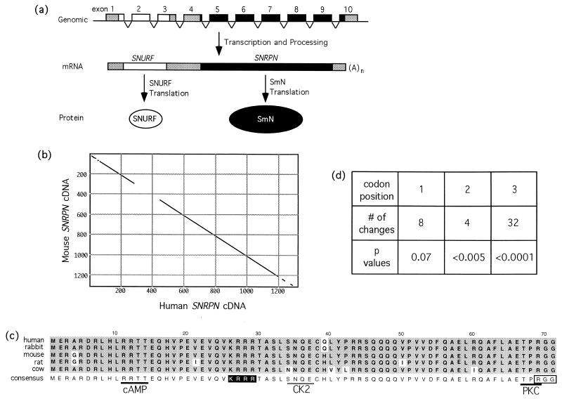
Polycistronic transcripts are common in prokaryotes but rare in eukaryotes. Phylogenetic analysis of the SNRPN (SmN) mRNA in five eutherian mammals reveals a second highly conserved coding sequence, termed SNURF (SNRPN upstream reading frame). The vast majority of nucleotide substitutions in SNURF occur in the wobble codon position, providing strong evolutionary evidence for selection for protein-coding function. Because SNURF-SNRPN maps to human chromosome 15q11-q13 and is paternally expressed, each cistron is a candidate for a role in the imprinted Prader-Willi syndrome (PWS) and PWS mouse models. SNURF encodes a highly basic 71-aa protein that is nuclear-localized (as is SmN). Because SNURF is the only protein-coding sequence within the imprinting regulatory region in 15q11-q13, it may have provided the original selection for imprinting in this domain. Whereas some human tissues express a minor SNURF-only transcript, mouse tissues express only the bicistronic Snurf-Snrpn transcript. We show that both SNURF and SNRPN are translated in normal, but not PWS, human, and mouse tissues and cell lines. These findings identify SNURF as a protein that is produced along with SmN from a bicistronic transcript; polycistronic mRNAs therefore are encoded in mammalian genomes where they may form functional operons.


Figures

Figure 1
Figure 1
SNURF–SNRPN gene structure and sequence analysis. (a) Schematic representation of the bicistronic SNURF–SNRPN locus depicting the progression from a contiguous nonoverlapping series of exons to a single transcript with two ORFs, to independent protein products. Open and filled structures correspond to SNURF or SNRPN, respectively, whereas the shaded regions indicate untranslated sequences. (b) Dot-plot analysis of full-length mouse and human SNRPN cDNAs; conserved sequences (window = 30, 65% minimum) appear as a diagonal line. (c) Amino acid alignment of putative SNURF proteins from five eutherian mammals. The derived consensus is shown below the compilation; residues consistent with the consensus are shaded in gray, changes are unshaded. Potential features appended to the consensus are: a nuclear localization signal (white letters on a black background), a C-terminal RGG motif (boxed), and phosphorylation sites that are absolutely conserved (heavy underline; cAMP, cAMP-dependent kinase; PKC, protein kinase C) or partially conserved (light underline; CK2, casein kinase II). (d) Codon position of the nucleotide changes that occur in the SNURF coding sequence of the five species examined. P values were derived from a χ2 analysis at each position.
Figure 2
Figure 2
SNURF-specific mRNA analyses. (a) Extended exposure of a human multitissue Northern blot probed with SNURF exons 1–3. A major 1.6-kb product is present in all tissues, and a minor 0.5-kb species is seen in a subset of tissues. Only the 1.6-kb product is seen with probes from SNRPN exons 4–10 (8). Tissue types are: H, heart; B, brain; Pl, placenta; Lu, lung; Li, liver; SM, skeletal muscle; K, kidney; Pa, pancreas. (b) SNURF exon 3b. The exon is shown as boxed, capital letters, with flanking genomic sequence shown as lower case. Consensus sequences for polyadenylation (white letters in a black box) and splice acceptor (splice consensus, conformity indicated by a vertical line; y, pyrimidine) also are shown. An alignment of the homologous mouse genomic sequence is shown below; ∗ marks a point mutation of the splice acceptor consensus in mice. (c) Extended exposure of a mouse multitissue Northern blot probed with mouse Snurf exons 1–3. The 1.6-kb product is present in most tissues, but a 0.5-kb band is not observed in any tissue. Identical results are obtained for probes from Snrpn exons 4–10 (data not shown). Tissue types are as for a. S, spleen; T, testis.
Figure 3
Figure 3
SNURF protein analyses. (a) Representative immunoprecipitation-Western analyses of human and mouse tissues with an ≈9-kDa SNURF form. N, normal lymphoblast; PWa, PWS109 lymphoblast line; ES, mouse embryonic stem cell line; WT, normal mouse brain; PW, Prader-Willi mouse model. (b) Immunoprecipitation–Western analysis of human muscle tissues with an ≈11-kDa SNURF isoform. N, normal heart and skeletal muscle from the same individual (no. 2,144); PWb, heart muscle from PWS individual (no. 1,199), PWc, skeletal muscle from PWS individual (no. 1,889). Lymphoblast designations are as in a. 293, human kidney 293 cells that have been transiently transfected with SNURF-(His)6 (+), or mock empty vector (−). (c) Intracellular localization of ectopically expressed underivatized GFP or GFP fused to the C terminus of SNURF.
Figure 4
Figure 4
Western analysis of SmN. (a) SmN in human lymphoblasts. N, normal; AS, AS139; PWS, PWS109. (b) SmN in mouse tissues. ES, embryonic stem cells; PW, murine PWS model brain extract; WT, normal mouse brain extract. SmA, SmN, SmB′, SmB, SmC, and SmD complex are indicated in the left margins of a and b.

References

    1. Bartolomei M S, Tilghman S M. Annu Rev Genet. 1997;31:493–525. - PubMed
    1. Cassidy S B. J Med Genet. 1997;34:917–923. - PMC - PubMed
    1. Nicholls R D, Saitoh S, Horsthemke B. Trends Genet. 1998;14:194–200. - PubMed
    1. Schmauss C, Brines M L, Lerner M R. J Biol Chem. 1992;267:8521–8529. - PubMed
    1. Sutcliffe J S, Nakao M, Christian S, Örstavik K H, Tommerup N, Ledbetter D H, Beaudet A L. Nat Genet. 1994;8:52–58. - PubMed

Publication types

MeSH terms