. 2000 May;182(9):2520-9.
doi: 10.1128/JB.182.9.2520-2529.2000.

The trimethylamine methyltransferase gene and multiple dimethylamine methyltransferase genes of Methanosarcina barkeri contain in-frame and read-through amber codons

Affiliations

The trimethylamine methyltransferase gene and multiple dimethylamine methyltransferase genes of Methanosarcina barkeri contain in-frame and read-through amber codons

L Paul et al. J Bacteriol. 2000 May.

Abstract

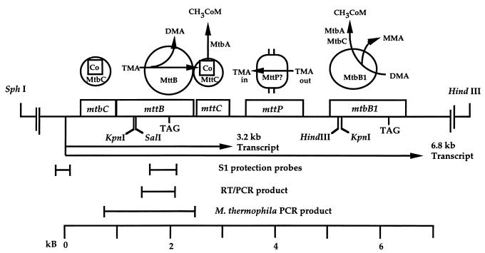
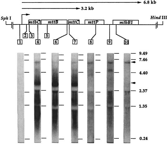
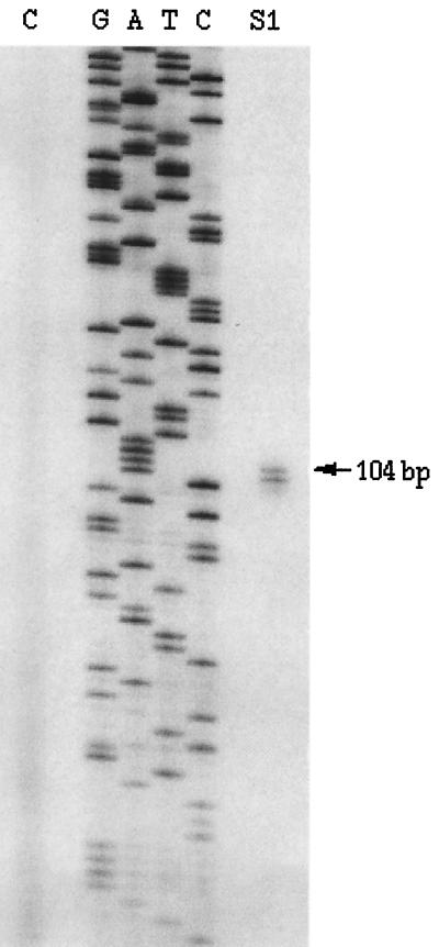
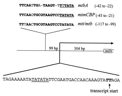
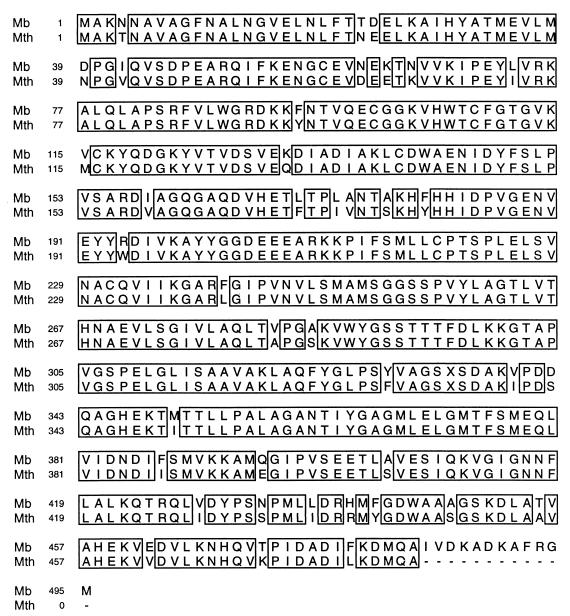
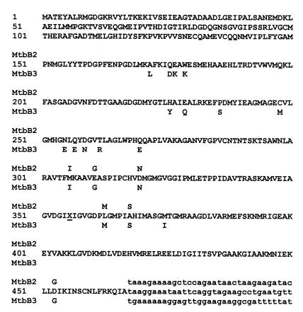
Three different methyltransferases initiate methanogenesis from trimethylamine (TMA), dimethylamine (DMA) or monomethylamine (MMA) by methylating different cognate corrinoid proteins that are subsequently used to methylate coenzyme M (CoM). Here, genes encoding the DMA and TMA methyltransferases are characterized for the first time. A single copy of mttB, the TMA methyltransferase gene, was cotranscribed with a copy of the DMA methyltransferase gene, mtbB1. However, two other nearly identical copies of mtbB1, designated mtbB2 and mtbB3, were also found in the genome. A 6.8-kb transcript was detected with probes to mttB and mtbB1, as well as to mtbC and mttC, encoding the cognate corrinoid proteins for DMA:CoM and TMA:CoM methyl transfer, respectively, and with probes to mttP, encoding a putative membrane protein which might function as a methylamine permease. These results indicate that these genes, found on the chromosome in the order mtbC, mttB, mttC, mttP, and mtbB1, form a single transcriptional unit. A transcriptional start site was detected 303 or 304 bp upstream of the translational start of mtbC. The MMA, DMA, and TMA methyltransferases are not homologs; however, like the MMA methyltransferase gene, the genes encoding the DMA and TMA methyltransferases each contain a single in-frame amber codon. Each of the three DMA methyltransferase gene copies from Methanosarcina barkeri contained an amber codon at the same position, followed by a downstream UAA or UGA codon. The C-terminal residues of DMA methyltransferase purified from TMA-grown cells matched the residues predicted for the gene products of mtbB1, mtbB2, or mtbB3 if termination occurred at the UAA or UGA codon rather than the in-frame amber codon. The mttB gene from Methanosarcina thermophila contained a UAG codon at the same position as the M. barkeri mttB gene. The UAG codon is also present in mttB transcripts. Thus, the genes encoding the three types of methyltransferases that initiate methanogenesis from methylamine contain in-frame amber codons that are suppressed during expression of the characterized methyltransferases.


Figures

FIG. 1
FIG. 1
Schematic of the mtt-mtb1 transcriptional unit. Above the gene sequence are indicated the reactions demonstrated for the gene products, i.e., the DMA and TMA methyltransferases and their cognate corrinoid proteins. The function of mttP is proposed but has not been demonstrated. The locations of probes used in S1 protection studies are shown along with the reverse transcriptase PCR product confirming the presence of the UAG codon within the mttB transcript. The M. thermophila mttB gene was amplified by PCR and corresponds to the region indicated above it. Key restriction sites used during cloning of the complete set of genes are indicated.
FIG. 2
FIG. 2
Three nearly identical copies of the DMA methyltransferase gene were cloned as HindIII fragments from M. barkeri MS and sequenced. In order to establish that mtbB1 is linked to the mtt genes, a restriction map of genomic DNA was made using Southern blots probed with oligonucleotides TCP10 and 18A (+4837 to +4852) (A). The map was compared with restriction sites sequenced in the 4.0-kb HindIII fragment (B), the 2.5-kb HindIII fragment (C), or the 2.0-kb HindIII fragment (D). Unsequenced DNA in the 2.5-kb HindIII fragment is indicated by the dashed line. Restriction sites are indicated as follows: Hd, HindIII; Hn, HincII; S, SalI; RI, EcoRI; D, DraI; RV, EcoRV; K, KpnI.
FIG. 3
FIG. 3
Northern blot analysis of transcripts produced from the DMA and TMA methyltransferase gene cluster. The hybridization targets for the probes are indicated by the lines connected to the boxed probe numbers above the resulting autoradiograms. The oligonucleotide probes were complementary to the following regions: 1, −97 to −115; 2, +181 to +196; 3, +396 to +413; 4, +833 to +848; 6, +2434 to +2464; 7, +2569 to +2591; 8, +4095 to +4114; 9, +4786 to +4800; and 10, +5215 to +5237. Probe 5 (+1299 to 1644) was a 5′-end-labeled fragment of mttB. Size standards are indicated to the right in kilobases. The migrations of standards, which varied slightly for different blots, are indicated by horizontal lines next to each blot.
FIG. 4
FIG. 4
Mapping of the mtt-mtb1 transcript start site by S1 analysis. Total cellular RNA was denatured and cooled in the presence of the radiolabeled restriction fragment shown in Fig. 1 and as described in Materials and Methods. The protected fragments of 103 and 104 nucleotides correspond to 303 and 304 bp from the translational start site of mtbC. The sequencing ladder was generated from pUC19. E. coli tRNA was used in place of M. barkeri RNA in the control lane (C).
FIG. 5
FIG. 5
Schematic of the upstream region and transcriptional start site of the mtt-mtb1 unit. In the lower part of the figure is the nucleotide sequence upstream and around the transcript start site. The first T of the two thymine bases identified by S1 mapping (boldface italics) is considered as the start of the transcript and is located 304 bases from the mtbC translation start site. As seen in the upper part of the figure, 99 bases upstream from the mtt-mtb1 transcript start site there is a highly conserved sequence in the regions 5′ of the mtmCBP and mtbA transcript start sites. The location of this sequence relative to the mapped start sites of these transcripts is indicated to the left of each sequence. The putative promoters for the mtbA, mtmCBP, and mtt-mtb1 transcripts are underlined.
FIG. 6
FIG. 6
Comparison of the deduced amino acid sequence of mttB amplified from M. thermophila (Mth) and mttB cloned from M. barkeri (Mb). The UAG codon position (at position 334) is indicated by X in both sequences. Identical residues are boxed.
FIG. 7
FIG. 7
Predicted gene products of available sequence from mtbB1, mtbB2, and mtbB3. The complete sequence of MtbB1 is shown and is numbered on the left-hand side. The first 48 residues shown for MtbB1 are not presently available for MtbB2 or MtbB3. The sequences of MtbB2 and MtbB3 are identical to that of MtbB1 for the next 100 residues; different residues that follow are indicated in the figure for MtbB2 or MtbB3 above and below, respectively, the MtbB1 sequence. The X at position 356 marks the UAG codon found in all three genes. The nucleotide sequence following the corresponding UAA or UGA codon of each gene is shown for comparison (lowercase).

References

    1. Altschul S F, Madden T L, Schaffer A A, Zhang J, Zhang Z, Miller W, Lipman D J. Gapped BLAST and PSI-BLAST: a new generation of protein database search programs. Nucleic Acids Res. 1997;25:3389–3402. - PMC - PubMed
    1. Böck A, Forchhammer K, Heider J, Leinfelder W, Sawers G, Veprek B, Zinoni F. Selenocysteine: the 21st amino acid. Mol Microbiol. 1991;5:515–520. - PubMed
    1. Boone D R, Whitman W B, Rouviére P. Diversity and taxonomy of methanogens. In: Ferry J G, editor. Methanogenesis. Ecology, physiology, biochemistry, and genetics. New York, N.Y: Chapman & Hall; 1993. pp. 35–80.
    1. Bult C J, White O, Olsen G J, Zhou L, Fleischmann R D, Sutton G G, Blake J A, FitzGerald L M, Clayton R A, Gocayne J D, Kerlavage A R, Dougherty B A, Tomb J F, Adams M D, Reich C I, Overbeek R, Kirkness E F, Weinstock K G, Merrick J M, Glodek A, Scott J L, Geoghagen N S M, Venter J C. Complete genome sequence of the methanogenic archaeon, Methanococcus jannaschii. Science. 1996;273:1058–1073. - PubMed
    1. Burke S A, Krzycki J A. Reconstitution of monomethylamine:coenzyme M methyl transfer with a corrinoid protein and two methyltransferases purified from Methanosarcina barkeri. J Biol Chem. 1997;272:16570–16577. - PubMed

Publication types

MeSH terms

Associated data

LinkOut - more resources