. 1999 Mar;181(6):1868-74.
doi: 10.1128/JB.181.6.1868-1874.1999.

Rapid hypothesis testing with Candida albicans through gene disruption with short homology regions

Affiliations

Rapid hypothesis testing with Candida albicans through gene disruption with short homology regions

R B Wilson et al. J Bacteriol. 1999 Mar.

Abstract

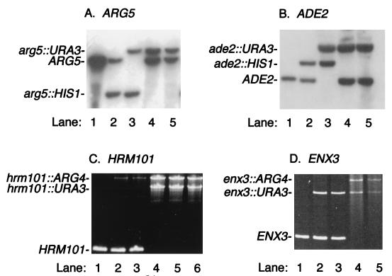
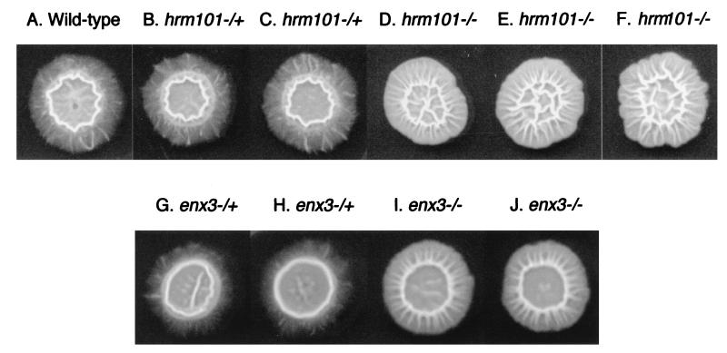
Disruption of newly identified genes in the pathogen Candida albicans is a vital step in determination of gene function. Several gene disruption methods described previously employ long regions of homology flanking a selectable marker. Here, we describe disruption of C. albicans genes with PCR products that have 50 to 60 bp of homology to a genomic sequence on each end of a selectable marker. We used the method to disrupt two known genes, ARG5 and ADE2, and two sequences newly identified through the Candida genome project, HRM101 and ENX3. HRM101 and ENX3 are homologous to genes in the conserved RIM101 (previously called RIM1) and PacC pathways of Saccharomyces cerevisiae and Aspergillus nidulans. We show that three independent hrm101/hrm101 mutants and two independent enx3/enx3 mutants are defective in filamentation on Spider medium. These observations argue that HRM101 and ENX3 sequences are indeed portions of genes and that the respective gene products have related functions.


Figures

FIG. 1
FIG. 1
Restriction maps. In panels A to E, thin horizontal lines represent DNA segments and thick lines represent the relevant open reading frame with 5′ and 3′ ends on the left and right, respectively. Panel D is drawn at a smaller scale than the other panels. Panels A to C represent the entire cassettes used for primer-directed gene disruption. (A) Insert of plasmid pGEM-URA3. The URA3 PCR product lies between the SphI and SalI sites in vector pGEM-T. (B) Insert of plasmid pGEM-HIS1. The HIS1 genomic fragment lies between the SphI and SalI sites in vector pGEM-T. (C) Insert of plasmid pRS-ARG4ΔSpeI. The ARG4 PCR product lies between the KpnI and SpeI sites in vector pRS314. (D) Genomic ARG5 locus. The open box represents the insertion/deletion allele arg5::hisG (12). Positions of the disruption primers and probe for Southern analysis are shown above the restriction map. (E) Genomic ADE2 locus. Positions of the disruption primers and probe for Southern analysis are shown above the restriction map. (F) Homology relationships between ARG5, the ARG5 disruption primers, and URA3 and HIS1 cassettes. The ARG5-5DR and ARG5-3DR primers are represented by arrows, which point to each primer’s 3′ end. Sets of parallel vertical lines indicate homology between the primers and ARG5 sequences or disruption cassette templates. The 461-bp segment of ARG5 shown above the primers is deleted from the arg5::hisG allele (12). This diagram is not to scale.
FIG. 2
FIG. 2
Schematic diagram of de novo ARG5 disruption. Each pair of parallel lines represents the two ARG5 alleles. Successive transformations are indicated by arrows. Strain RM1000 was transformed with an arg5::HIS1 PCR product; 4 of 24 transformants were arg5::HIS1/ARG5 heterozygotes, as indicated by Southern analysis. One heterozygote was transformed with an arg5::URA3 PCR product; 1 of 27 transformants was an arg5::HIS1/arg5::URA3 homozygous mutant, as indicated by Southern analysis and its Arg phenotype. Twenty-one of the remaining transformants were arg5::URA3/ARG5 heterozygotes, as indicated by their Arg+ His phenotype and, for a representative group, by Southern analysis.
FIG. 3
FIG. 3
Analysis of transformant strains. (A) Southern blot analysis of ARG5 disruption experiment. Genomic DNA samples, digested with BamHI, were prepared from strain RM1000 (ARG5/ARG5 [lane 1]), Arg-het1 (arg5::HIS1/ARG5 [lane 2]), a Uri+ His+ Arg transformant from Arg-het1 (arg5::HIS1/arg5::URA3 [lane 3]), and two Uri+ His Arg+ transformants from Arg-het1 (arg5::URA3/ARG5 [lanes 4 and 5]). The blot was probed with a 0.7-kbp ClaI-ScaI fragment of ARG5, as indicated in Fig. 1D. (B) Southern blot analysis of ADE2 disruption experiment. Genomic DNA samples, digested with BglII and HindIII, were prepared from strain RM1000 (ADE2/ADE2 [lane 1]), Ade-het1 (ade2::HIS1/ADE2 [lane 2]), a Uri+ His+ Ade (red) transformant from Ade-het1 (ade2::HIS1/ade2::URA3 [lane 3]), and two Uri+ His Ade+ transformants from Ade-het1 (ade2::URA3/ADE2 [lanes 4 and 5]). The blot was probed with a 0.8-kbp EcoRI-EcoRV fragment of ADE2, as indicated in Fig. 1E. (C) PCR analysis of HRM101 disruption experiment. PCR amplification with primers RIM101-5a and seq7 was carried out on genomic DNA samples from strain BWP17 (HRM101/HRM101 [lane 1]), Hrm-het1 (hrm1::ARG4/HRM101 [lane 2]), Hrm-het2 (hrm1::ARG4/HRM101 [lane 3]), and Uri+ Arg+ transformants from Hrm-het1 and Hrm-het2 (hrm1::ARG4/hrm101::URA3 [strains BWP29, BWP30, and BWP31] [lanes 4 to 6]). (D) PCR analysis of ENX3 disruption experiment. PCR amplification with primers PalA5′ and PalA3′ was carried out on genomic DNA samples from strain BWP17 (ENX3/ENX3 [lane 1]), Enx-het1 (enx3::URA3/ENX3 [lane 2]), Enx-het2 (enx3::URA3/ENX3 [lane 3]), and two Uri+ Arg+ transformants from Enx-het1 (enx3::URA3/enx3::ARG4 [strains DAY23 and DAY24] [lanes 4 and 5]).
FIG. 4
FIG. 4
Filamentation on Spider plates. Colonies were incubated for 6 days on Spider plates at 37°C. Strains were BWP17 (wild type [A]), Hrm-het1 and Hrm-het2 (both hrm1::ARG4/HRM101 [B and C]), BWP29, BWP30, and BWP31 (all hrm1::ARG4/hrm101::URA3 [D to F]), Enx-het1 and Enx-het2 (both enx3::URA3/ENX3 [G and H]), and DAY23 and DAY24 (both enx3::URA3/enx3::ARG4 [I and J]).

References

    1. Baudin A, Ozier-Kalogeropoulos O, Denouel A, Lacroute F, Cullin C. A simple and efficient method for direct gene deletion in Saccharomyces cerevisiae. Nucleic Acids Res. 1993;21:3329–3330. - PMC - PubMed
    1. Birse C E, Irwin M Y, Fonzi W A, Sypherd P S. Cloning and characterization of ECE1, a gene expressed in association with cell elongation of the dimorphic pathogen Candida albicans. Infect Immun. 1993;61:3648–3655. - PMC - PubMed
    1. Braun B R, Johnson A D. Control of filament formation in Candida albicans by the transcriptional repressor TUP1. Science. 1997;277:105–109. - PubMed
    1. Candida albicans information. 19 December 1998, revision date. [Online.] University of Minnesota, St. Paul. http://alces.med.umn.edu/Candida.html. [8 February 1999, last date accessed.]
    1. Candida tblastn search. 21 October 1998, revision date. [Online.] Stanford University, Palo Alto, Calif. http://candida.stanford.edu/btComb.html. [8 February 1999, last date accessed.]

Publication types

Associated data

LinkOut - more resources