. 2000 Sep;20(17):6568-78.
doi: 10.1128/MCB.20.17.6568-6578.2000.

Divergent N-terminal sequences target an inducible testis deubiquitinating enzyme to distinct subcellular structures

Affiliations

Divergent N-terminal sequences target an inducible testis deubiquitinating enzyme to distinct subcellular structures

H Lin et al. Mol Cell Biol. 2000 Sep.

Abstract

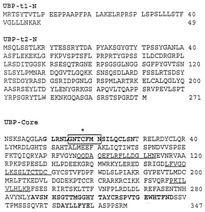
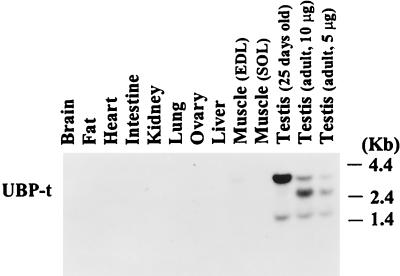
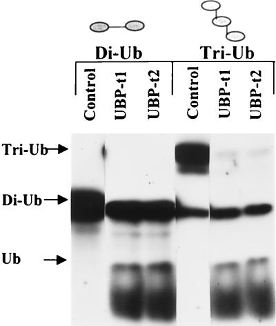
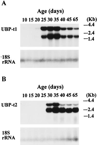
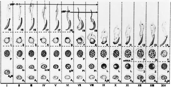
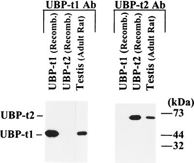
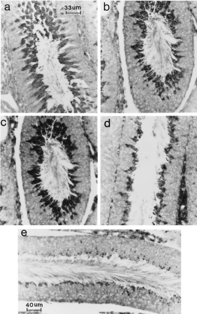
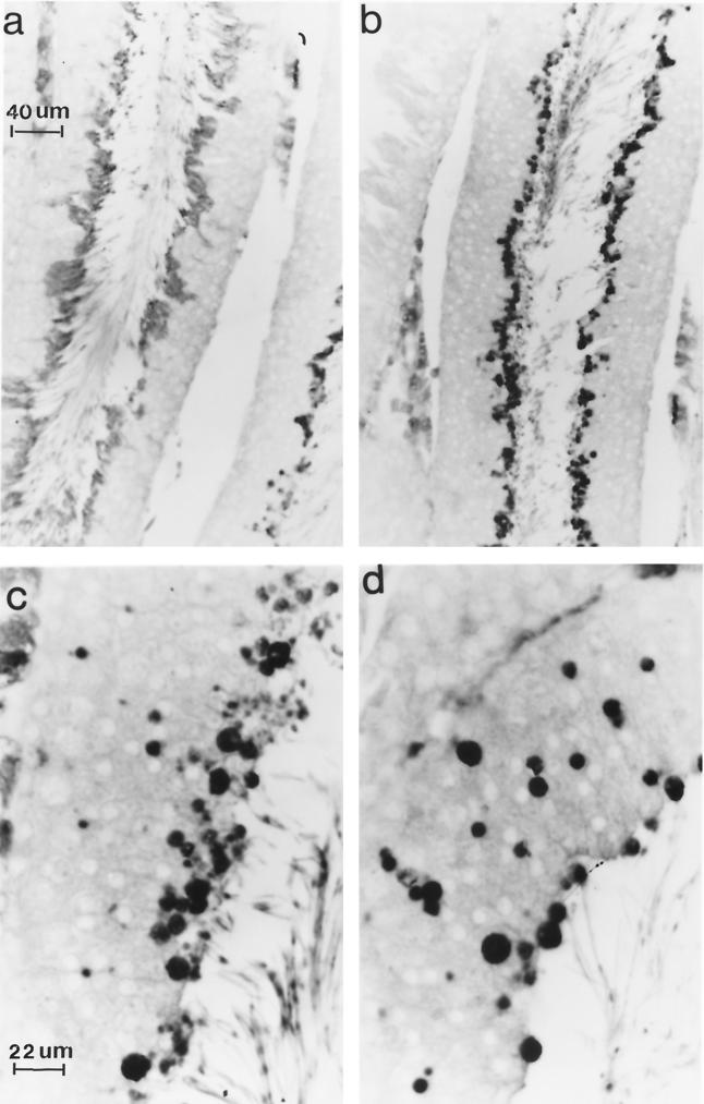
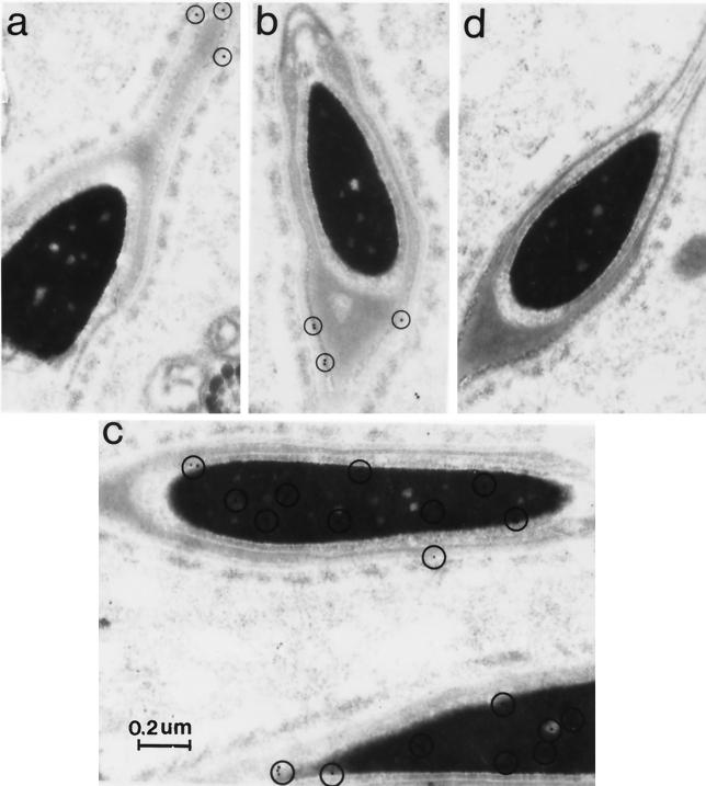
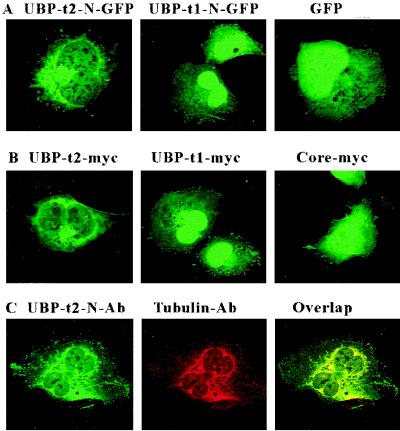
Ubiquitin-specific processing proteases (UBPs) presently form the largest enzyme family in the ubiquitin system, characterized by a core region containing conserved motifs surrounded by divergent sequences, most commonly at the N-terminal end. The functions of these divergent sequences remain unclear. We identified two isoforms of a novel testis-specific UBP, UBP-t1 and UBP-t2, which contain identical core regions but distinct N termini, thereby permitting dissection of the functions of these two regions. Both isoforms were germ cell specific and developmentally regulated. Immunocytochemistry revealed that UBP-t1 was induced in step 16 to 19 spermatids while UBP-t2 was expressed in step 18 to 19 spermatids. Immunoelectron microscopy showed that UBP-t1 was found in the nucleus while UBP-t2 was extranuclear and was found in residual bodies. For the first time, we show that the differential subcellular localization was due to the distinct N-terminal sequences. When transfected into COS-7 cells, the core region was expressed throughout the cell but the UBP-t1 and UBP-t2 isoforms were concentrated in the nucleus and the perinuclear region, respectively. Fusions of each N-terminal end with green fluorescent protein yielded the same subcellular localization as the native proteins, indicating that the N-terminal ends were sufficient for determining differential localization. Interestingly, UBP-t2 colocalized with anti-gamma-tubulin immunoreactivity, indicating that like several other components of the ubiquitin system, a deubiquitinating enzyme is associated with the centrosome. Regulated expression and alternative N termini can confer specificity of UBP function by restricting its temporal and spatial loci of action.


Figures

FIG. 1
FIG. 1
UBP-t1 and UBP-t2 sequences. Translation of cDNA clones encoding this testis-deubiquitinating enzyme revealed the presence of two isoforms, UBP-t1 and UBP-t2. UBP-t1-N indicates the amino-terminal sequence of the short isoform, and UBP-t2-N indicates the amino-terminal sequence of the long isoform. UBP-Core indicates the common sequence of both isoforms. The asterisk indicates an active-site cysteine; bold residues indicate the Cys and His motifs; other regions conserved in UBP family are underlined; residues in the box indicate the peptide sequence encoded by the degenerate oligonucleotide used in the 3′ RACE reaction to identify this cDNA.
FIG. 2
FIG. 2
Expression of UBP mRNA in rat tissues. RNA samples (10 μg) from the indicated tissues of adult (unless otherwise indicated) rats were resolved by electrophoresis on 1% agarose gels and transferred to a nylon membrane. Following hybridization with a 32P-labeled fragment from the UBP-t core region cDNA, the membrane was washed and exposed to film. EDL, extensor digitorum longus; SOL, soleus.
FIG. 3
FIG. 3
UBP-t1 and UBP-t2 have deubiquitinating-enzyme activities. Enzymatic activities toward 125I-diubiquitin (Di-Ub) joined in a peptide bond and 125I-triubiquitin polymer (Tri-Ub) linked via lysine 48 isopeptide bonds were measured in lysates prepared from E. coli cells expressing either UBP-t1 or UBP-t2. Reaction products were separated by SDS-PAGE and detected by autoradiography. An equivalent volume of bacterial lysate not expressing UBP-t1 or UBP-t2 was used as the negative control.
FIG. 4
FIG. 4
Induction of UBP-t1 and UBP-t2 mRNAs during postnatal development of the testis. RNA (10 μg) from rat testes of different ages were electrophoresed on a 1% agarose gel and transferred to a nylon membrane. After hybridization with cDNA probes derived from the 5′ end of UBP-t1 (A) or UBP-t2 (B), the blots were subjected to autoradiography. Following removal of the probe, the membranes were rehybridized with a probe based on the 18S rRNA to evaluate loading and transfer of the samples to the membrane.
FIG. 5
FIG. 5
The 14 cellular associations or stages observed in the seminiferous epithelium of the rat. Each vertical column, depicted by a Roman numeral, represents a cellular association and shows the various cell types present at that stage. The stage of the cycle is identified by means of 14 of the 19 steps of spermiogenesis (numbers 1 to 19). These steps are defined by the changes observed in the nucleus and acrosomal system in semithin sections (0.5 μm thick) stained with toluidine blue. The cellular associations or stages of the cycle succeed one another in time in any given area of the seminiferous epithelium according to the sequence indicated from left to right in the figure. Following stage XIV, stage I reappears, so that the sequence starts over again. The succession of the 14 stages makes up the cycle of the seminiferous epithelium. The mitotic divisions of the spermatogonia are indicated by the letter M. The germ cells present are A1, A2, A3, and A4 (type A spermatogonia); In (intermediate-type spermatogonia); B (type B spermatogonia); P1 (preleptotene primary spermatocytes); L (leptotene primary spermatocytes); Z (zygotene primary spermatocytes); P (pachytene primary spermatocytes); Di (diplotene primary spermatocytes); II (secondary spermatocytes); and 1 to 19 (steps of spermiogenesis). The dotted line indicates the cell types which express UBP-t mRNA, as determined by in situ hybridization (data not shown). Solid lines indicate cell types expressing UBP-t1 and UBP-t2 proteins. Modified from reference and reproduced with permission of the publisher.
FIG. 6
FIG. 6
UBP-t1 and UBP-t2 antibodies are specific. Bacterial lysates containing the full length of UBP-t1 or UBP-t2 or the homogenates of 65-day-old rat testes were resolved by SDS-PAGE on 10% polyacrylamide gels and transferred to nitrocellulose membranes. The membranes were probed with anti-UBP-t1 N-terminal extension or UBP-t2 N-terminal extension antibody separately and then incubated with horseradish peroxidase-conjugated protein A and subjected to chemiluminescent detection.
FIG. 7
FIG. 7
UBP-t1 protein is expressed in step 16 to 19 spermatids. A cross section of rat seminiferous epithelium was immunostained with anti-UBP-t1 N-terminus-specific antibody and reacted with a peroxidase-conjugated anti-rabbit IgG. The reaction was visualized under a light microscope. (a) A cross section of seminiferous tubule at stage III of the cycle of the seminiferous epithelium demonstrates that the reaction is over step 16 spermatids. (b) A cross section of seminiferous tubule at stage V of the cycle of the seminiferous epithelium shows that the reaction is over step 17 spermatids. (c) A cross section of seminiferous tubule at stage VI of the cycle of the seminiferous epithelium shows that the reaction is over step 18 spermatids. (d) A cross section of seminiferous tubule at stage VII of the cycle of the seminiferous epithelium demonstrates that the reaction is over step 19 spermatids. (e) A cross section of seminiferous tubule at stage VIII of the cycle of the seminiferous epithelium shows that the reaction is over step 19 spermatids. Residual bodies were not stained but appeared dark because a blue filter used during photography did not eliminate the methylene blue counterstain. (Preincubation of antibody with GST fused to the UBP-t1 N terminus before hybridization with the sections resulted in the absence of any staining [data not shown]). Magnification in panels b, c, and d is the same as in panel e.
FIG. 8
FIG. 8
UBP-t2 protein is expressed in step 18 to 19 spermatids. A cross section of rat seminiferous epithelium was immunostained with anti-UBP-t2 N-terminus-specific antibody and reacted with a peroxidase-conjugated anti-rabbit IgG. The reaction was visualized under a light microscope. (a) A cross section of the seminiferous tubule at stage VI of the cycle of the seminiferous epithelium demonstrates that the reaction is over step 18 spermatids. (b) A cross section of the seminiferous tubule at stage VIII of the cycle of the seminiferous epithelium shows that the reaction is over step 19 spermatids. (c) A cross section of the seminiferous tubule at stage VIII of the cycle of the seminiferous epithelium shows that the reaction is over step 19 spermatids and residual bodies. (d) A cross section of the seminiferous tubule at stage VIII of the cycle of the seminiferous epithelium shows that the reaction is over residual bodies. (Preincubation of antibody with GST fused to the UBP-t2 N terminus before hybridization with the sections resulted in the absence of any staining [data not shown]). Magnification in panel b is the same as in panel a; magnification of panel d is the same as in panel c.
FIG. 9
FIG. 9
UBP-t1 and UBP-t2 are differentially distributed in spermatids. Rat testis ultrathin sections were mounted on Formvar-coated nickel grids, stained with anti-UBP-t1 or anti-UBP-t2 specific antibody, and incubated with colloidal gold-conjugated goat anti-rabbit antibody. Sections were counterstained with uranyl acetate followed by lead citrate. As a negative control, the anti-UBP-t1 or anti-UBP-t2 antibody was preincubated with excess GST–UBP-t1 or GST–UBP-t2 protein for 2 h at 37°C prior to use on the sections. Electron micrographs were taken on a Philips 400 electron microscope. The circled small black dots represent positive staining. (a and b) Anti-UBP-t2-specific antibody staining. (c) Anti-UBP-t1-specific antibody staining. (d) Negative control.
FIG. 10
FIG. 10
N-terminal sequences of UBP-t1 and UBP-t2 determine subcellular localization. (A) COS-7 cells were transfected with plasmids expressing either the UBP-t1 N-terminal extension (UBP-t1-N-GFP) or the UBP-t2 N-terminal extension (UBP-t2-N-GFP) fused to GFP or the GFP alone. Cells were fixed and examined using a confocal fluorescence microscope. (B) COS-7 cells were transfected with plasmids expressing myc epitope tags of either UBP-t1, UBP-t2, or the core region alone. Following fixation, the cells were stained sequentially with anti-myc antibody and FITC-conjugated goat anti-mouse IgG antibody and examined with a confocal fluorescence microscope. (C) COS-7 cells were transfected with a plasmid expressing myc-tagged UBP-t2. Following fixation, the cells were stained with both anti-amino-terminal extension of UBP-t2 polyclonal antibody and anti-γ-tubulin monoclonal antibody followed by both FITC-conjugated goat anti-rabbit IgG antibody (green) and TRITC-conjugated goat anti-mouse IgG antibody (red). Confocal microscopy was used to detect anti-UBP-t2 fluorescence (UBP-t2-N-Ab) and anti-γ-tubulin fluorescence (Tubulin-Ab) and to analyze for colocalizing signals (Overlap, yellow).

References

    1. Amerik A, Swaminathan S, Krantz B A, Wilkinson K D, Hochstrasser M. In vivo disassembly of free polyubiquitin chains by yeast Ubp14 modulates rates of protein degradation by the proteasome. EMBO J. 1997;16:4826–4838. - PMC - PubMed
    1. Anton L C, Schubert U, Bacik I, Princiotta M F, Wearsch P A, Gibbs J, Day P M, Realini C, Rechsteiner M C, Bennink J R, Yewdell J W. Intracellular localization of proteasomal degradation of a viral antigen. J Cell Biol. 1999;146:113–124. - PMC - PubMed
    1. Baker R T, Tobias J W, Varshavsky A. Ubiquitin-specific proteases of Saccharomyces cerevisiae. Cloning of UBP2 and UBP3, and functional analysis of the UBP gene family. J Biol Chem. 1992;267:23364–23375. - PubMed
    1. Clermont Y, Oko R, Hermo L. Cell biology of mammalian spermiogenesis. In: Desjardins C, editor. Cell and molecular biology of the testis. New York, N.Y: Oxford University Press; 1993. pp. 332–375.
    1. Coux O, Tanaka K, Goldberg A L. Structure and functions of the 20S and 26S proteasomes. Annu Rev Biochem. 1996;65:801–847. - PubMed

Publication types

MeSH terms

Associated data

LinkOut - more resources