. 2000 Jun 6;97(12):6820-5.
doi: 10.1073/pnas.97.12.6820.

Human monoclonal antibodies reactive to oligodendrocytes promote remyelination in a model of multiple sclerosis

Affiliations

Human monoclonal antibodies reactive to oligodendrocytes promote remyelination in a model of multiple sclerosis

A E Warrington et al. Proc Natl Acad Sci U S A. .

Abstract

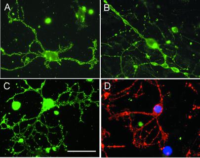
Promoting remyelination, a major goal of an effective treatment for demyelinating diseases, has the potential to protect vulnerable axons, increase conduction velocity, and improve neurologic deficits. Strategies to promote remyelination have focused on transplanting oligodendrocytes (OLs) or recruiting endogenous myelinating cells with trophic factors. Ig-based therapies, routinely used to treat a variety of neurological and autoimmune diseases, underlie our approach to enhance remyelination. We isolated two human mAbs directed against OL surface antigens that promoted significant remyelination in a virus-mediated model of multiple sclerosis. Four additional OL-binding human mAbs did not promote remyelination. Both human mAbs were as effective as human i.v. Ig, a treatment shown to have efficacy in multiple sclerosis, and bound to the surface of human OLs suggesting a direct effect of the mAbs on the cells responsible for myelination. Alternatively, targeting human mAbs to areas of central nervous system (CNS) pathology may facilitate the opsonization of myelin debris, allowing repair to proceed. Human mAbs were isolated from the sera of individuals with a form of monoclonal gammopathy. These individuals carry a high level of monoclonal protein in their blood without detriment, lending support to the belief that administration of these mAbs as a therapy would be safe. Our results are (i) consistent with the hypothesis that CNS-reactive mAbs, part of the normal Ig repertoire in humans, may help repair and protect the CNS from pathogenic immune injury, and (ii) further challenge the premise that Abs that bind OLs are necessarily pathogenic.


Figures

Figure 1
Figure 1
The methodology used to quantify white matter, white matter pathology, and remyelination in the spinal cords of TMEV-infected mice. Light photomicrograph of a thoracic level spinal cord section from an SJL/J mouse chronically infected with TMEV and treated with polyclonal human IgM (A). White matter at the periphery stains darker than the lighter central gray matter. The area of total white matter is traced (indicated by the red outlines), at a magnification of ×40. Then at a magnification of ×100 the areas of white matter pathology are traced (indicated by the green outlines). In this example, the areas of white matter pathology appear as lighter areas at the periphery of the section. Finally, at a magnification of ×250 the areas of OL remyelination (indicated by the blue outlines) and SC remyelination (indicated by the yellow outline) are traced. OL remyelination is characterized by thin myelin sheaths in relation to axon diameter. The percent area of white matter pathology is calculated by dividing the area in green by the area in red × 100. The percent area of OL remyelination is calculated by dividing the area in blue by the area in green × 100. Ten spinal cord cross sections are traced for each animal considered and the areas are combined to calculate a score for that animal. Generally, 7–8 animals are treated in each experimental group to allow for deaths and animals that did not contain at least 5% total white matter pathology. Usually 4–5 treated animals met the criteria for inclusion into the final data set. A high magnification field of the dorsal column white matter (B, from the area indicated by * in A) demonstrates significant OL remyelination (arrow). (Scale bars are 250 μm in A and 20 μm in B.)
Figure 2
Figure 2
After treatment with human Abs, chronically TMEV-infected mice demonstrate significant OL remyelination. Light photomicrographs of representative areas of spinal cord white matter pathology of different treatment groups. Treatment with IVIg resulted in significant OL remyelination (A). Almost complete OL remyelination, characterized by densely packed thin myelin sheaths in relation to axon diameter (B, arrowhead), was observed in sections from the spinal cords of mice after treatment with polyclonal human IgM (B) and human mAbs sHIgM 22 (F) and sHIgM46 (G). In contrast, after treatment with human mAbs sHIgM1 (C), sHIgM2 (D), sHIgM14 (E), or PBS (H) mice demonstrated white matter pathology without significant OL remyelination. Infiltrating inflammatory cells, macrophages ingesting myelin debris (A, arrowhead), and signs of active myelin destruction were also evident. Spinal cord cross sections in four of eight animals treated with sHIgM22 and five of five animals treated with sHIgM46 contained at least one area of nearly confluent OL remyelination, a rare event indicating significant tissue repair. In contrast, the 10 spinal cord cross sections from each mouse treated with sHIgM1, sHIgM2, sHIgM14, or PBS contained none. (Scale bar is 20 μm.)
Figure 3
Figure 3
Human mAbs isolated for their ability to bind to rat OLs also bind to the surface of human OLs in culture. sHIgM14 (A), which did not promote remyelination, and sHIgM22 (B) and sHIgM46 (C), which did promote remyelination, bound to the perikaryon and elaborate process and membrane extensions of sulfatide-positive human OLs maintained in culture for 3 weeks. sHIgM2 (D, green channel) is an example of a human mAb that did not bind to sulfatide positive (D, red channel) human OLs. Nuclei are labeled blue. IVIg, polyclonal human IgM, and human mAbs sHIgM1 and sHIgM2 did not bind to the surface of human OLs at any time point examined. (Scale bar is 25 μm.)

References

    1. Perier O, Gregoire A. Brain. 1965;88:937–952. - PubMed
    1. Prineas J W, Connel F. Ann Neurol. 1979;5:22–31. - PubMed
    1. Wolswijk G, Nobel M. Development (Cambridge, UK) 1989;105:387–400. - PubMed
    1. Armstrong R C, Dorn H H, Kufta C V, Friedman E, Dubois-Dalcq M E. J Neurosci. 1992;12:1538–1547. - PMC - PubMed
    1. Gumpel M, Baumann N, Raoul M, Jaques C. Neurosci Lett. 1983;37:307–312. - PubMed

Publication types

MeSH terms

Associated data