. 2000 Oct 10;97(21):11439-44.
doi: 10.1073/pnas.200360997.

Cloning of a type I cytokine receptor most related to the IL-2 receptor beta chain

Affiliations

Cloning of a type I cytokine receptor most related to the IL-2 receptor beta chain

K Ozaki et al. Proc Natl Acad Sci U S A. .

Abstract

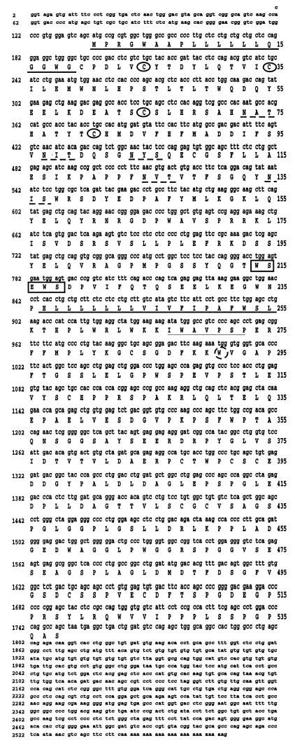
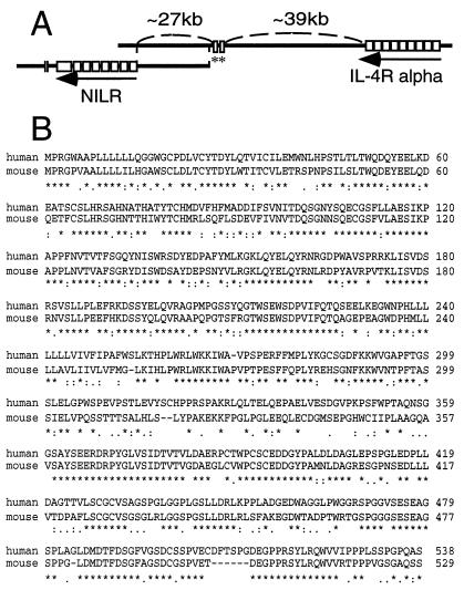
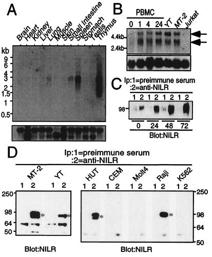
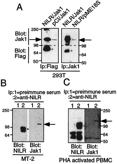
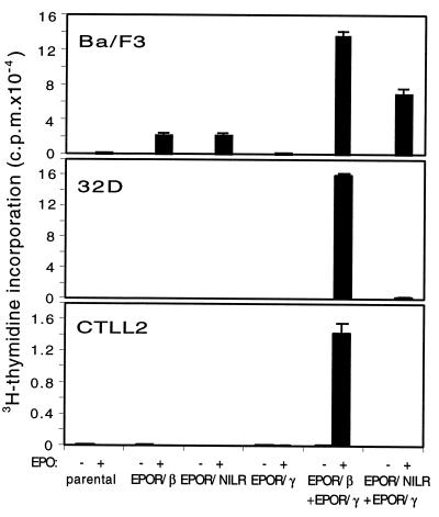
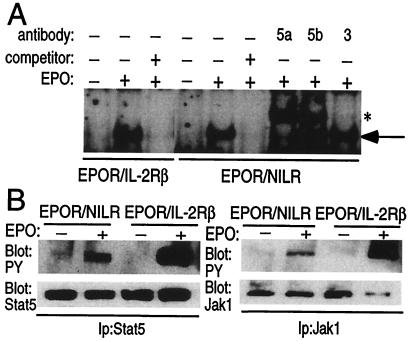
We have identified a type I cytokine receptor, which we have termed novel interleukin receptor (NILR), that is most related to the IL-2 receptor beta chain (IL-2Rbeta) and physically adjacent to the IL-4 receptor alpha chain gene on chromosome 16. NILR mRNA is most highly expressed in thymus and spleen, and is induced by phytohemagglutinin in human peripheral blood mononuclear cells. NILR protein was detected on human T cell lymphotropic virus type I-transformed T cell lines, Raji B cells, and YT natural killer-like cells. Artificial homodimerization of the NILR cytoplasmic domain confers proliferation to Ba/F3 murine pro-B cells but not to 32D myeloid progenitor cells or CTLL-2 murine helper T cells. In these latter cells, heterodimerization of IL-2Rbeta and the common cytokine receptor gamma chain (gamma(c)) cytoplasmic domains allows potent proliferation, whereas such heterodimerization of NILR with gamma(c) does not. This finding suggests that NILR has signaling potential but that a full understanding of its signaling partner(s) is not yet clear. Like IL-2Rbeta, NILR associates with Jak1 and mediates Stat5 activation.


Figures

Figure 1
Figure 1
NILR encodes a type I cytokine receptor. DNA and predicted amino acid sequences of human NILR cDNA clone 17. Conserved Cys residues are circled. The predicted signal peptide and transmembrane domains are underlined. The boundaries of the transmembrane domain are as predicted by ref. ; tmpred instead predicts that the domain begins at Leu-238 instead of at His-237. The potential N-linked glycosylation sites are underlined with broken lines. The WSXWS motif is boxed. Thin underline and broken circles denote the Box B1 region and conserved Trp, respectively.
Figure 2
Figure 2
The NILR and IL4RA genes are adjacent, and human and murine NILR genes are highly related. (A) Schematic showing two overlapping BAC clones that together span the NILR gene. The BAC clone positioned to the left contains all NILR coding exons; the one on the right contains at least two noncoding exons of NILR (asterisks) and the IL4RA gene. Marked distances are approximate. (B) Alignment of deduced human and murine NILR amino acid sequences by using clustal w (11). Asterisks, colons, and periods indicate identical amino acids and conservative and semiconservative substitutions, respectively.
Figure 3
Figure 3
NILR mRNA and protein expression. (A) (Upper). NILR mRNA expression distribution in mouse tissue. (Lower) Hybridization with a control human β-actin probe. (B) NILR mRNA expression is induced by PHA in human PBMC. Cells were stimulated with PHA for the indicated times and poly(A)+ RNA was isolated. NILR mRNA expression was also evaluated in YT (a positive control because YT cells were the source of the cDNA library), MT-2, and Jurkat T cell lines. Arrows indicate NILR mRNA. (Lower) Hybridization with the pHe7 probe as a control (12). (C) NILR protein is induced by PHA in human PBMC. Cells were stimulated with PHA for the indicated time periods, and preimmune serum (lane 1) or anti-NILR antiserum (lane 2) immunoprecipitates were blotted with anti-NILR antiserum. For each immunoprecipitation, 3.0 × 10⁷ cells were used. (D) NILR protein is expressed in MT-2, YT, HUT-102B2, and Raji cells, but not in CEM, Molt4, or K562 cells. Immunoprecipitates of preimmune serum (lane 1) or anti-NILR antiserum (lane 2) were blotted with anti-NILR antiserum. For each immunoprecipitation, 2.0–2.5 × 10⁷ cells were used.
Figure 4
Figure 4
Association of Jak1 with NILR. (A) FLAG-tagged NILR and Jak1 associate in transfected 293T cells. Indicated plasmids were transiently transfected. Samples were immunoprecipitated with anti-FLAG (Left) or anti-Jak1 (Right). The upper part of the membrane (>98 kDa) was blotted with anti-Jak1 and the lower part (<98 kDa) was blotted with anti-FLAG. Bands corresponding to NILR and Jak1 are indicated by asterisks and arrows, respectively. (B and C) Anti-NILR immunoprecipitates from HTVL-I-transformed MT-2 cells (B) and PHA-activated PBMC (C) contain Jak1. Preimmune serum (lane 1) or anti-NILR (lane 2) immunoprecipitates were divided; one part was blotted with anti-NILR antiserum (Left) and the other one was blotted with anti-Jak1 (Right). The asterisk and arrow indicate NILR and Jak1, respectively.
Figure 5
Figure 5
The NILR cytoplasmic domain can transduce a proliferative signal in Ba/F3 but not 32D and CTLL-2 cells. Ba/F3, 32D, and CTLL-2 cells expressing the indicated chimeric receptors were incubated with medium (open bars) or EPO at 100 units/ml (filled bars). After 92 h, 1 μCi of [3H]thymidine was added and cells were harvested 4 h later. As expected, Ba/F3 and 32D transfectants proliferated well in response to WEHI-3B-conditioned medium and CTLL-2 transfectants proliferated well in response to IL-2 (data not shown).
Figure 6
Figure 6
Electrophoretic mobility-shift assays and anti-phosphotyrosine immunoblotting of Ba/F3 cells expressing the indicated chimeric receptor. (A) Cells maintained in EPO (0.5 units/ml) were deprived of growth factor and then stimulated or not stimulated with EPO (20 units/ml, 30 min). Nuclear extracts were then prepared and binding assays were performed. The arrow indicates a DNA-binding protein corresponding to Stat5 and the asterisk indicates the bands that are detected after supershifting with antibodies to either murine Stat5a or Stat5b. (B) Cells were deprived of growth factor and then stimulated or not stimulated with EPO (20 units/ml, 10 min). Stat5 and Jak1 immunoprecipitates were blotted with anti-Stat5, anti-Jak1, or anti-phosphotyrosine antibodies as indicated.

References

    1. Leonard W J, O'Shea J J. Annu Rev Immunol. 1998;16:293–322. - PubMed
    1. Leonard W J. In: Fundamental Immunology. Paul W E, editor. Philadelphia: Lippincott; 1999. pp. 741–774.
    1. Miyajima A, Mui A L, Ogorochi T, Sakamaki K. Blood. 1993;82:1960–1974. - PubMed
    1. Leonard W J, Shores E W, Love P E. Immunol Rev. 1995;148:97–114. - PubMed
    1. Takeshita T, Asao H, Ohtani K, Ishii N, Kumaki S, Tanaka N, Munakata H, Nakamura M, Sugamura K. Science. 1992;257:379–382. - PubMed

Associated data