. 2002 Jul 1;22(13):5412-22.
doi: 10.1523/JNEUROSCI.22-13-05412.2002.

Serotonin stimulates phosphorylation of Aplysia synapsin and alters its subcellular distribution in sensory neurons

Affiliations

Serotonin stimulates phosphorylation of Aplysia synapsin and alters its subcellular distribution in sensory neurons

Annie Angers et al. J Neurosci. .

Abstract

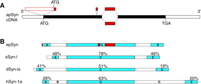
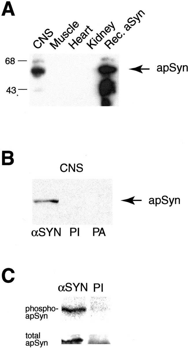
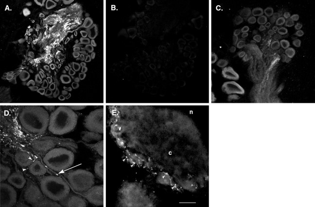
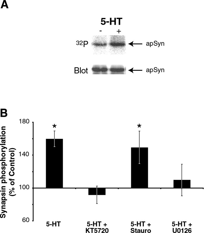
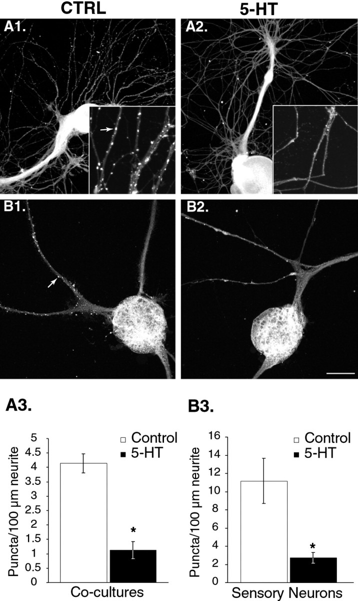
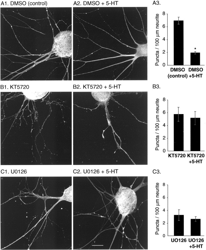
Only a small fraction of neurotransmitter-containing synaptic vesicles (SVs), the readily releasable pool, is available for fast Ca(2+)-induced release at any synapse. Most SVs are sequestered at sites away from the plasma membrane and cannot be exocytosed directly. Recruitment of SVs to the releasable pool is thought to be an important component of short-term synaptic facilitation by serotonin (5-HT) at Aplysia sensorimotor synapses. Synapsins are associated with SVs and hypothesized to play a central role in the regulation of SV mobilization in nerve terminals. Aplysia synapsin was cloned to examine its role in synaptic plasticity at the well characterized sensorimotor neuron synapse of this animal. Acute 5-HT treatment of ganglia induced synapsin phosphorylation. Immunohistochemical analyses of cultured Aplysia neurons revealed that synapsin is distributed in distinct puncta in the neurites. These puncta are rapidly dispersed after treatment of the neurons with 5-HT. The dispersion of synapsin puncta by 5-HT was fully reversible after washout of the modulator. Both 5-HT-induced phosphorylation and dispersion of synapsin were mediated, at least in part, by cAMP-dependent protein kinase and mitogen-activated protein kinase. These experiments indicate that synapsin and its regulation by 5-HT may play an important role in the modulation of SV trafficking in short-term synaptic plasticity.


Figures

Fig. 1.
Fig. 1.
Cloning of Aplysia synapsins (apSyn). A, Schematic representation of the synapsin cDNAs. The open boxes represent the untranslated regions, and the filled boxes represent the open reading frame. Alternative exons are represented above and below their site of insertion. B, Comparison of the domain structure and homology of Aplysia synapsin protein with squid, Drosophila, and human synapsin Ia. The degree of sequence identity between Aplysia and other synapsins is indicated above each identified domain. Variable regions in apSyn are shown in red.
Fig. 2.
Fig. 2.
Western blot analysis and antibody specificity.A, A polyclonal antibody raised against rec-apSyn protein recognizes a 57 kDa protein in CNS extracts. The lower molecular weight bands in the rec-apSyn lane are probably degradation products of the purified protein. The antibody also reacted strongly against rec-apSyn. The protein was not detectable in tissue extracts of heart, muscle, or kidney. B, The specificity of the antibody is demonstrated by blotting the same CNS extract with preimmune serum (PI) or the antisera preabsorbed with rec-aSyn (PA). C, The αSyn antibody can specifically immunoprecipitate a band of ∼57 kDa from CNS extracts (αSyn lane), whereas the preimmune serum cannot (PI lane). The band can be visualized by32P labeling (top panel) or immunoblotting with αSyn antibody (bottom panel).
Fig. 3.
Fig. 3.
apSyn immunofluorescence in sections of the pleural-pedal or abdominal ganglia. A, Low magnification of a pedal ganglion section showing abundant staining in the neuropil. Neuron somata are also lightly stained. No staining was observed in the surrounding connective tissue. B, Very low level of nonspecific signal is observed in an adjacent section incubated with preimmune serum. C, ApSyn immunofluorescence in a section of a pleural ganglion. The most intense signal is observed in the neuropil. Neuronal somata are also immunoreactive with a lower amount of signal. D, Higher magnification of the section in C showing the punctate nature of the signal in neurites. The arrow points to a varicosity-like region containing intense immunoreactivity. Arrowheads show similar structures on the surface of a cell body. E, High-power magnification of a large neuron from a left caudal section of an abdominal ganglion. Many brightly stained varicosities can be observed on small neurites running on the surface of the cell body (arrowheads). Cytoplasm (c) and nucleus (n) are visible. Scale bar:A, B, 150 μm; C, 75 μm; D, E, 25 μm.
Fig. 4.
Fig. 4.
Phosphorylation of apSyn. A, Immunoprecipitation of 32P-labeled apSyn (top) shows that apSyn is phosphorylated after 5 min treatment with 5-HT. The immunoblot on the bottom panelshows that equivalent amounts of apSyn were immunoprecipitated from control and treated ganglia. B, Summary data showing quantification of apSyn phosphorylation. Results are presented as the ratio of the 32P signal in the treated ganglia and control ganglia. Five minute treatment with 5-HT induces significant phosphorylation of apSyn (n = 6). One hour pretreatment with the PKA inhibitor KT5720 (n = 3) or the MAPK inhibitor U0126 (n = 6) inhibits 5-HT-induced phosphorylation of apSyn. Pretreatment with the PKC inhibitor staurosporine (n = 7) did not inhibit phosphorylation of apSyn by 5-HT. The asterisk indicates significant difference (p < 0.05) from controls.
Fig. 5.
Fig. 5.
Treatment with 5-HT modifies subcellular distribution of apSyn. A1, ApSyn immunoreactivity in cocultures is enriched in small puncta along neurites (Ctrl). A2, Five minute treatment with 5-HT (50 μm bolus, 1 μm final bath concentration) induces a dramatic dissipation of apSyn puncta. Theinset shows a higher magnification of the neurites. Thearrow points to a punctum. B1, Similar puncta were observed in isolated sensory neurons in culture (arrow). B2, After treatment with 5-HT, very few puncta could be observed along the neurites of the cells.A3, B3, Summary data presenting the number of puncta counted per 100 μm of measured neurites in cocultures (n = 6 for controls and 4 for 5-HT) (A3), and isolated sensory neurons (n = 6 for controls and 5 for 5-HT) (B3). Treatment of the cells with 5-HT before fixation dramatically reduced the number of puncta. Scale bar: A,B, 75 μm; insets, 25 μm.
Fig. 6.
Fig. 6.
VAMP distribution is not altered by 5-HT.A1, Under resting conditions, the subcellular distribution of VAMP appears punctate and similar to synapsin (compare with Fig. 5B). Arrowheads point to VAMP puncta (scale bar, 25 μm). A2, Treatment with 5-HT (5 min, 10 μm) does not alter VAMP localization, suggesting that short-term treatment with 5-HT does not significantly affect the organization of SV pools. B, Summary data illustrating the lack of effect of 5-HT on the density of VAMP puncta (n = 5 for controls and 5 for 5-HT).
Fig. 7.
Fig. 7.
Distribution of apSyn puncta after low-frequency stimulation and 5-HT-induced facilitation. A, Averaged amplitude of EPSPs during a train of 25 stimuli (○,n = 3) or with 5-HT application at the 10th stimuli of the train (●, n = 4). Note the increase of the average amplitude of the EPSPs beginning ∼4 sec after the application of 5-HT. B1, ApSyn immunoreactivity in cocultures is enriched in small puncta along the neurites of cells that received a train of 25 stimuli (Stim.). B2, Treatment with 5-HT during the train of stimuli (Stim. + 5-HT) induces dispersion of apSyn puncta. In bothB1 and B2, cells were fixed for subsequent immunohistochemistry as fast as possible after the 25th stimulus, <30 sec after exposure to 5-HT. The insetsshow a higher magnification of the neurites. The arrowin B1 points to an apSyn punctum. Scale bar:B1, B2, 75 μm; insets, 25 μm. C, Summary data presenting the number of puncta counted per 100 μm of measured neurites in control cocultures that received a train of 25 stimuli (Stim.) and cocultures that received the train of stimuli and were treated with 5-HT (Stim. + 5-HT).
Fig. 8.
Fig. 8.
Time course of the effect of 5-HT treatment on apSyn subcellular distribution in isolated SNs. A, Application of 5-HT for 5 min (A2) induces a dramatic dissipation of apSyn puncta compared with control conditions (A1). Very few apSyn puncta can be observed 15 min after removal of 5-HT (A3), but the protein distribution returned to control levels by 2 hr after removal of the modulator (A4). Scale bar, 25 μm. B, Summary data presenting the number of puncta counted per 100 μm of neurites. Treatment of the cells with 5-HT before fixation dramatically reduced the number of puncta, which returned to control level by 2 hr after termination of the treatment (A1,n = 5; A2, n = 5; A3, n = 5; A4,n = 4).
Fig. 9.
Fig. 9.
ApSyn subcellular redistribution induced by treatment with 5-HT is inhibited by the PKA inhibitor KT5720 and the MAPK cascade inhibitor U0126.A, DMSO (vehicle for inhibitors) did not affect the ability of 5-HT to induce redistribution of apSyn in cultured SNs after a 5 min incubation period (A3, DMSO:n = 14; DMSO + 5-HT:n = 12). B, One hour treatment with the PKA inhibitor KT5720 completely inhibited the ability of 5-HT to induce redistribution of apSyn (B3,KT5720: n = 12; KT5720 + 5-HT: n = 11). C, The MEK inhibitor U0126 also appeared to preclude the effects of 5-HT, although the number of puncta before 5-HT application was altered by the presence of the inhibitor (C3, U0126:n = 9; U0126 + 5-HT:n = 9). Scale bar, 25 μm.

References

    1. Angers A, Bean AJ, Byrne JH. Cloning and molecular characterization of Aplysia synaptic vesicle protein synapsin. Soc Neurosci Abstr. 1999;25:1749.
    1. Angers A, Chin J, Cleary LJ, Byrne JH. 5-HT treatment induces redistribution of Aplysia synapsin. Soc Neurosci Abstr. 2000;26:881.
    1. Bähler M, Greengard P. Synapsin I bundles F-actin in a phosphorylation-dependent manner. Nature. 1987;326:704–707. - PubMed
    1. Benfenati F, Bahler M, Jahn R, Greengard P. Interactions of synapsin I with small synaptic vesicles: distinct sites in synapsin I bind to vesicle phospholipids and vesicle proteins. J Cell Biol. 1989;108:1863–1872. - PMC - PubMed
    1. Benfenati F, Valtorta F, Chieregatti E, Greengard P. Interaction of free and synaptic vesicle-bound synapsin I with F-actin. Neuron. 1992;8:377–386. - PubMed

Publication types

MeSH terms

Associated data

LinkOut - more resources