. 2002 Apr;128(4):1200-11.
doi: 10.1104/pp.010928.

Steroleosin, a sterol-binding dehydrogenase in seed oil bodies

Affiliations

Steroleosin, a sterol-binding dehydrogenase in seed oil bodies

Li-Jen Lin et al. Plant Physiol. 2002 Apr.

Erratum in

  • Plant Physiol 2002 Aug;129(4):1930

Abstract

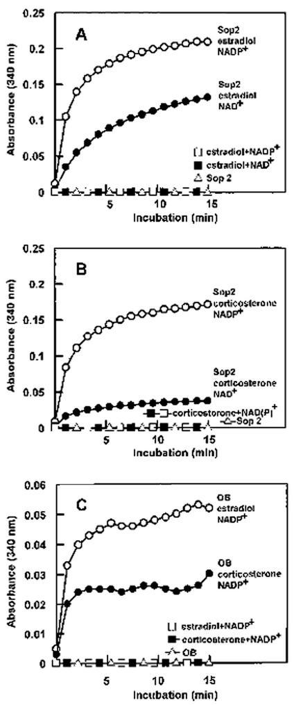
Besides abundant oleosin, three minor proteins, Sop 1, 2, and 3, are present in sesame (Sesamum indicum) oil bodies. The gene encoding Sop1, named caleosin for its calcium-binding capacity, has recently been cloned. In this study, Sop2 gene was obtained by immunoscreening, and it was subsequently confirmed by amino acid partial sequencing and immunological recognition of its overexpressed protein in Escherichia coli. Immunological cross recognition implies that Sop2 exists in seed oil bodies of diverse species. Along with oleosin and caleosin genes, Sop2 gene was transcribed in maturing seeds where oil bodies are actively assembled. Sequence analysis reveals that Sop2, tentatively named steroleosin, possesses a hydrophobic anchoring segment preceding a soluble domain homologous to sterol-binding dehydrogenases/reductases involved in signal transduction in diverse organisms. Three-dimensional structure of the soluble domain was predicted via homology modeling. The structure forms a seven-stranded parallel beta-sheet with the active site, S-(12X)-Y-(3X)-K, between an NADPH and a sterol-binding subdomain. Sterol-coupling dehydrogenase activity was demonstrated in the overexpressed soluble domain of steroleosin as well as in purified oil bodies. Southern hybridization suggests that one steroleosin gene and certain homologous genes may be present in the sesame genome. Comparably, eight hypothetical steroleosin-like proteins are present in the Arabidopsis genome with a conserved NADPH-binding subdomain, but a divergent sterol-binding subdomain. It is indicated that steroleosin-like proteins may represent a class of dehydrogenases/reductases that are involved in plant signal transduction regulated by various sterols.


Figures

Figure 1
Figure 1
Sequence alignment of sesame and Arabidopsis Sop2 sequences. The sequences are aligned according to four proposed structural regions (oil body-anchoring segment, NADPH binding subdomain, active site, and sterol-binding subdomain) of Sop2. The amino acid number for the last residue in each row is listed on the right for each species. A gap represented by a broken line is introduced between residues 241 (Ala) and 242 (Gly) of sesame Sop2 for best alignment. Three partial sequences obtained directly from amino acid sequencing are boxes. The three consensus residues in the active site are highlighted. Predicted secondary structures are indicated on the tops of the sequences (see Fig. 5B for details). The locations of α-helices and β-strands in the predicted Sop2 structure are indicated and are labeled successively. Locations where introns in their corresponding genomic sequences occur are indicated by triangles on tops of the sequences. The accession number of the aligned Arabidopsis Sop2 is BAA96983.
Figure 2
Figure 2
SDS-PAGE and western blotting of the overexpressed sesame Sop2 in E. coli. Along with sesame oil body proteins and purified Sop2, the recombinant Sop2 (without oil body-anchoring segment) overexpressed in E. coli using a His-tag fusion vector was resolved in a 12.5% (w/v) SDS-PAGE gel. A duplicate gel was transferred onto nitrocellulose membrane and was then subjected to immunodetection using antibodies (1:1,500 dilution) against the seed-purified Sop2 protein. Labels on the left indicate the molecular masses of proteins.
Figure 3
Figure 3
SDS-PAGE and western blotting of proteins extracted from seed oil bodies of various species. Proteins extracted from oil bodies of sesame and three other oily seeds were resolved in a 10% (w/v) SDS-PAGE gel. A duplicate gel was transferred onto nitrocellulose membrane and was then subjected to immunoassaying using antibodies (1:100 dilution) against sesame Sop2. Labels on the left indicate the molecular masses of proteins.
Figure 4
Figure 4
Northern-blot analysis of total RNA extracted from various stages of maturing sesame seeds. Each lane was loaded with 20 μg of total RNA extracted from maturing seeds at various days after flowering (DAF). After blotting, the membrane was hybridized with a 32P-labeled probe containing the coding sequence of sesame Sop2, caleosin, or oleosin. Only the portion of the membrane corresponding to the visible hybridized RNA is shown.
Figure 5
Figure 5
Sequence alignment of sesame Sop2 with six sterol-binding dehydrogenase/reductase sequences. Sesame Sop2 is compared with different sterol-binding dehydrogenases/reductases of diverse species. The amino acid number for the last residue in each row is listed on the right for each species. Broken lines in the sequences represent gaps introduced for best alignment and conserved residues are shaded. The proposed structural regions (membrane anchoring, NADPH binding, and sterol binding) are indicated on the tops of the sequences. The three consensus residues in dehydrogenase/reductase active site are highlighted. The accession numbers of the aligned sequences are: Human-1 (Homo sapiens), AAC31757; Droso-1 (Drosophila melanogaster), AAF56927; Human-2, AAF06941; Droso-2, AAF45573; Strepto (Streptomyces clavuligerus), AAF86624; Bacillus (Bacillus subtilis), CAB14310.
Figure 6
Figure 6
A, Hydropathy plot of sesame Sop2. The hydrophobicity scale was plotted versus amino acid sequence of sesame Sop2 with a window size of 19 using hydropathy index described by Kyte and Doolittle (1982). B, A secondary structural model of sesame steroleosin on the surface of an oil body. A monolayer of PLs, depicted by pink balls attached with two tails, segregates the hydrophobic TAG matrix (gradient yellow) of an oil body from hydrophilic cytosol (gradient light blue). Amino acid residues are represented by one-letter symbols in green circles. Numbers next to residues or secondary structures represent their relative positions counting from N terminus. Two structural domains are predicted in a steroleosin molecule: an N-terminal oil body-anchoring domain and a sterol-binding dehydrogenase domain. The hydrophobic N-terminal domain (residues 1–40) is supposed to associate with the monolayer PL of oil body surface by forming two amphipathic α-helices connected by a hydrophobic segment termed the Pro knob. The core structure of sterol-binding dehydrogenase domain forms a seven-stranded β-sheet surrounded by α-helices and can be divided into three regions: an NADPH-binding subdomain, an active site, and a sterol-binding subdomain. The three conserved residues (S, Y, and K) in the active site are indicated. NADPH and sterol are denoted by brown and orange molecules, respectively. C, Three-dimensional structural modeling of the sterol-binding dehydrogenase domain of sesame steroleosin. The three-dimensional structure of the soluble domain comprising the core structure of sterol-binding dehydrogenase was predicted using homology modeling (Lund et al., 1997).
Figure 7
Figure 7
Spectrophotometric detection of dehydrogenase activity in sesame steroleosin (Sop2). The dehydrogenase activity of the expressed Sop2 soluble domain (25 μg) was detected using estradiol (A) or corticosterone (B) as a sterol substrate in the presence of NADP+ or NAD+. C, Sesame oil bodies (OB) containing 5 μg of Sop2 protein was assayed for dehydrogenase activity using the same sterol substrates in the presence of NADP+.
Figure 8
Figure 8
Southern-blot analysis of genomic DNA extracted from leaves of sesame plants. Each lane was loaded with 10 μg of genomic DNA completely digested with EcoRI, HindIII, or PstI. After blotting, the membrane was hybridized with a 32P-labeled probe containing part of the coding sequence of sesame steroleosin.
Figure 9
Figure 9
Sequence alignment of eight putative sterol-binding dehydrogenase/reductase sequences in Arabidopsis. The amino acid number for the last residue in each row is listed on the right for each species. Broken lines in the sequences represent gaps introduced for best alignment and conserved residues are shaded. The proposed structural regions (N-terminal appendix, NADPH binding, and sterol binding) are indicated on the tops of the sequences. The three consensus residues in the dehydrogenase/reductase active site are highlighted. Locations where introns in their corresponding genomic sequences occur are indicated by triangles on the tops of the sequences. The accession numbers of Arab-1 through -8 sequences are: BAA96983, BAA96990, BAB09144, BAA96982, CAB51207, CAB51208, CAB39626, and AAF01606.

References

    1. Abell BM, Holbrook LA, Abenes M, Murphy DJ, Hills MJ, Moloney MM. Role of the proline knot motif in oleosin endoplasmic reticulum topology and oil body targeting. Plant Cell. 1997;9:1481–1493. - PMC - PubMed
    1. Altschul SF, Warren G, Webb M, Eugene WM, David JL. Basic local alignment search tool. J Mol Biol. 1990;215:403–410. - PubMed
    1. Branden CI. Relation between structure and function of α/β proteins. Q Rev Biophys. 1980;13:317–338. - PubMed
    1. Breton R, Housset D, Mazza C, Fontecilla-Camps JC. The structure of a complex of human 17β-hydroxysteroid dehydrogenase with estradiol and NADP+identifies two principal targets for the design of inhibitors. Structure. 1996;15:905–915. - PubMed
    1. Chen ECF, Tai SSK, Peng CC, Tzen JTC. Identification of three novel unique proteins in seed oil bodies of sesame. Plant Cell Physiol. 1998;39:935–941. - PubMed

Publication types

MeSH terms

Associated data

LinkOut - more resources