. 2005 Mar;71(3):1531-8.
doi: 10.1128/AEM.71.3.1531-1538.2005.

Two alpha(1-3) glucan synthases with different functions in Aspergillus fumigatus

Affiliations

Two alpha(1-3) glucan synthases with different functions in Aspergillus fumigatus

A Beauvais et al. Appl Environ Microbiol. 2005 Mar.

Abstract

Alpha(1-3) glucan is a main component of the Aspergillus fumigatus cell wall. In spite of its importance, synthesis of this amorphous polymer has not been investigated to date. Two genes in A. fumigatus, AGS1 and AGS2, are highly homologous to the AGS genes of Schizosaccharomyces pombe, which encode putative alpha(1-3) glucan synthases. The predicted Ags proteins of A. fumigatus have an estimated molecular mass of 270 kDa. AGS1 and AGS2 were disrupted in A. fumigatus. Both Deltaags mutants have similar altered hyphal morphologies and reduced conidiation levels. Only Deltaags1 presented a reduction in the alpha(1-3) glucan content of the cell wall. These results showed that Ags1p and Ags2p were functionally different. The cellular localization of the two proteins was in agreement with their different functions: Ags1p was localized at the periphery of the cell in connection with the cell wall, whereas Ags2p was intracellularly located. An original experimental model of invasive aspergillosis based on mixed infection and quantitative PCR was developed to analyze the virulence of A. fumigatus mutant and wild-type strains. Using this model, it was shown that the cell wall and morphogenesis defects of Deltaags1 and Deltaags2 were not associated with a reduction in virulence in either mutant. This result showed that a 50% reduction in the content of the cell wall alpha(1-3) glucan does not play a significant role in A. fumigatus pathogenicity.


Figures

FIG. 1.
FIG. 1.
Box-shade representation of the amino acid sequence similarities of the Ags proteins of A. fumigatus (Af) and S. pombe (Sp). AfAgs1p, aa 1321 to 1582; AfAgs2p, aa 1316 to 1575; SpAgs1p, aa 1300 to 1558; SpAgs2p, aa 1302 to 1556; SpAgs3p, aa 1328 to 1585; SpAgs4p, aa 1308 to 1566; and Sp Ags5p, aa 411 to 667 (note that this protein lacks the N-terminal region). Amino acid sequences used for degenerate oligonucleotide primers are bracketed.
FIG. 2.
FIG. 2.
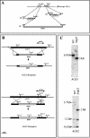
Characterization of Δags mutants. (A) Construction of the knockout plasmid pΔags1. (1) First integration of the HindIII-XhoI fragment of ags1DisC in pBluscript SK(+) digested at HindIII and XhoI; (2) second integration of the NotI-EcoRV fragment of ags1DisN in Sk(+)-ags1DisC digested at NotI and EcoRV; and (3) third integration of the StuI-HindIII fragment of the HPH fragment in Sk(+)-ags1DisC-ags1DisN digested at EcoRV and HindIII. The same strategy was used for the knockout plasmid pΔags2. (B) Schematic representation showing the predicted disruption events resulting from homologous integration of the AfAGS1 and AfAGS2 deletion constructs into the A. fumigatus genome. (C) Southern blot analysis of Δags1 and Δags2 mutants compared to the WT. Genomic DNA was digested with NcoI and hybridized with ags1DisC or ags2DisC, labeled “probe” in panel B.
FIG. 3.
FIG. 3.
Hyphal morphology of Δags2 and WT strains grown for 16 h in Brian’s medium at 25°C. Note the dichotomous branching of the apex of Δags2 (a) compared to the normal apical growth of the WT (b). Magnification, ×1,400.
FIG. 4.
FIG. 4.
Conidiation of Δags1 and Δags2 mutants and the parental WT strain of A. fumigatus after 1 week of growth on malt medium at 25°C. (A) Bar, standard error of the mean calculated for at least three replicates. (B) A. fumigatus head and phialides of WT (a), Δags2 (b), and Δags1 (c). Magnification, ×2,800.
FIG. 5.
FIG. 5.
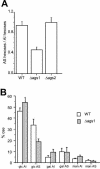
(A) Total hexose content of the AS and AI fractions for the parental strain (WT) and the Δags1 and Δags2 mutants. The hexose concentration was estimated as milligram equivalents of glucose. (B) Comparison of the monosaccharide composition of AS and AI fractions of the cell wall from parental (WT) and Δags1. glc, glucose; gal, galactose; man, mannose. As an example, the value for glucose in the AI fraction was calculated as:  formula imagewhere [glcAI] is the concentration in milligram equivalents of glucose in the AI fraction. Bar, standard error of the mean calculated for at least three replicates.
FIG. 6.
FIG. 6.
Immunolocalization of Ags1p and Ags2p in germ tubes of A. fumigatus WT, using anti-Agsp antiserum or preimmune serum and goat anti-rabbit-fluorescein isothiocyanate-conjugated antibodies. A TCS4D Leica confocal laser scanning microscope was used, and the same exposure time was used for control and assay experiments. Bar, 5 μm. (A) Control preimmune serum of two different germinated conidia (panels 1 and 2 and 3 and 4, respectively). (B) Immunofluorescence microscopy of two different germinated conidia (panels 1 and 2 and 3 and 4, respectively) using anti-Ags1p antibody. (C) Immunofluorescence microscopy of two different germinated conidia (panels 1 and 2 and 3 and 4, respectively) using anti-Ags2p antibody. A1, A3, B1, B3, C1, and C3 are shown by phase-contrast light; A2, A4, B2, B4, C2, and C4 are shown by epifluorescence.

References

    1. Beauvais, A., and J. P. Latgé. 2001. Membrane and cell wall targets in Aspergillus fumigatus. Drug Resist. Updates. 4:1-12. - PubMed
    1. Beauvais, A., J. M. Bruneau, P. C. Mol, M. J. Buitrago, R. Legrand, and J. P. Latgé. 2001. Glucan synthase complex of Aspergillus fumigatus. J. Bacteriol. 183:2273-2279. - PMC - PubMed
    1. Brian, P. W., A. W. Dawkins, J. F. Grove, H. G. Hemming, D. Lowe, and G. L. Norris. 1961. Phytotoxic compounds produced by Fusarium equiseti. J. Exp. Bot. 12:1-7.
    1. Claros, M. G., and G. van Heijne. 1994. TopRed II: an improved software for membrane protein structure predictions. Comput. Appl. Biol. Sci. 10:685-686. - PubMed
    1. Dijkgraaf, G. J. P., M. Abe, Y. Ohya, and H. Bussey. 2002. Mutations in Fks1p affect the cell wall content of β-1,3- and β-1,6-glucan in Saccharomyces cerevisiae. Yeast 19:671-690. - PubMed

Associated data

LinkOut - more resources