. 2001 Apr 24;98(9):5222-7.
doi: 10.1073/pnas.081075398. Epub 2001 Apr 17.

Characterization of a Plasmodium falciparum erythrocyte-binding protein paralogous to EBA-175

Affiliations

Characterization of a Plasmodium falciparum erythrocyte-binding protein paralogous to EBA-175

D C Mayer et al. Proc Natl Acad Sci U S A. .

Abstract

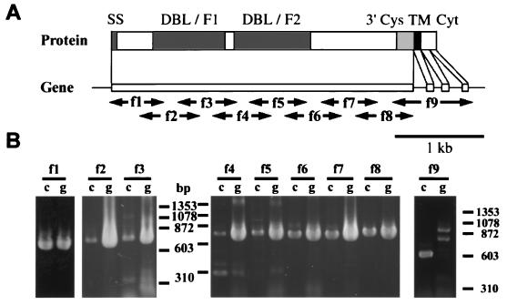
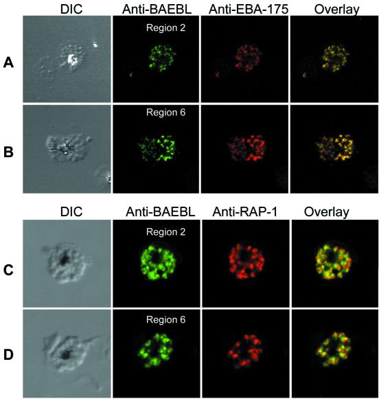
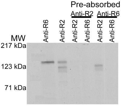
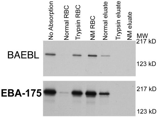
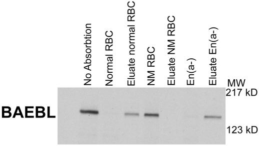
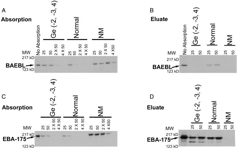
A member of a Plasmodium receptor family for erythrocyte invasion was identified on chromosome 13 from the Plasmodium falciparum genome sequence of the Sanger Centre (Cambridge, U.K.). The protein (named BAEBL) has homology to EBA-175, a P. falciparum receptor that binds specifically to sialic acid and the peptide backbone of glycophorin A on erythrocytes. Both EBA-175 and BAEBL localize to the micronemes, organelles at the invasive ends of the parasites that contain other members of the family. Like EBA-175, the erythrocyte receptor for BAEBL is destroyed by neuraminidase and trypsin, indicating that the erythrocyte receptor is a sialoglycoprotein. Its specificity, however, differs from that of EBA-175 in that BAEBL can bind to erythrocytes that lack glycophorin A, the receptor for EBA-175. It has reduced binding to erythrocytes with the Gerbich mutation found in another erythrocyte, sialoglycoprotein (glycophorin C/D). The interest in BAEBL's reduced binding to Gerbich erythrocytes derives from the high frequency of the Gerbich phenotype in some regions of Papua New Guinea where P. falciparum is hyperendemic.


Figures

Figure 1
Figure 1
Sequencing strategy and exon/intron structure of baebl. Oligonucleotides were designed based on the genomic sequence obtained from the P. falciparum genome project (Sanger Centre) and used for the sequencing of genomic DNA (GenBank Accession No. AF332918) and reverse transcription–PCR of mRNA (GenBank Accession No. AF332919) to determine the intron/exon structure in P. falciparum Dd2/Nm strain. (A) Schematic representation of the gene and predicted protein structure of baebl. Predicted protein structure has strong similarity with EBA-175, containing the putative signal sequence (SS, amino acids 1–21) predicted by SIGNALP V2.0; region 2 (two DBL domains, F1 and F2); region 6 (3′Cys), the transmembrane domain (TM, amino acids 1,134–1,153) predicted by TMHMM V2.0, followed by the putative cytoplasmic domain (Cyt). (B) f1 to f9 primers (see Materials and Methods) are used for reverse transcription–PCR of mRNA (lanes marked c) and PCR of genomic DNA (lanes marked g). (Bar = 1 kb.)
Figure 2
Figure 2
Confocal microscopy demonstrates the localization of BAEBL in micronemes. (A) Dd2/Nm schizonts were double labeled with anti-BAEBL region 2 and anti-EBA-175. Schizonts immunolabeled with anti-BAEBL region 2 were stained with Alexa 488 secondary antibody (green). Schizonts labeled with anti-EBA-175 were stained with Alexa 594 secondary antibody (red). (B) Dd2/Nm schizonts were double labeled with anti-BAEBL region 6 and anti-EBA-175. Schizonts immunolabeled with anti-BAEBL region 6 were stained with Alexa 488 secondary antibody (green). Schizonts labeled with anti-EBA-175 were stained with Alexa 594 secondary antibody (red). (C) Dd2/Nm schizonts were double labeled with anti-BAEBL region 2 and anti-RAP-1 monoclonal antibody. Schizonts immunolabeled anti-BAEBL region 2 were stained with Alexa 488 secondary antibody (green). Schizonts labeled with anti-RAP-1 were stained with tetramethyl rhodamine isothiocyanate (TRITC) secondary antibody (red). (D) Dd2/Nm schizonts were double labeled with anti-BAEBL region 6 and anti-RAP-1 monoclonal antibody. Schizonts immunolabeled with anti-BAEBL region 6 were stained with Alexa 488 secondary antibody (green). Schizonts labeled with anti-RAP-1 were stained with TRITC secondary antibody (red).
Figure 3
Figure 3
Evidence that anti-region 2 (Anti-R2) and anti-region 6 (Anti-R6) sera immunoprecipitate the same protein. The supernatant was preabsorbed with either anti-region 2 or anti-region 6 followed by immunoprecipitation by the two sera. BAEBL was removed by both sera. MW, molecular mass.
Figure 4
Figure 4
BAEBL and EBA-175 did not bind to neuraminidase (NM eluate) or trypsin-treated erythrocytes (Trypsin RBC). Eluates of BAEBL and EBA-175 were seen only from normal erythrocytes.
Figure 5
Figure 5
BAEBL binds and is eluted from En(a−) erythrocytes that lack glycophorin A. NM RBC are neuraminidase-treated normal erythrocytes. MW, molecular mass; kD, kilodalton.
Figure 6
Figure 6
Absorption and elution of BAEBL (A and B) and EBA-175 (C and D) with various amounts (25, 50, 2 × 50, and 4 × 50 μl of packed erythrocytes) of Gerbich [Ge(−2, −3, 4)], normal, and neuraminidase (NM)-treated erythrocytes. For elution, 25 and 50 μl of packed erythrocytes were used. MW, molecular mass; kD, kilodalton.

References

    1. Dvorak J A, Miller L H, Whitehouse W C, Shiroishi T. Science. 1975;187:748–750. - PubMed
    1. Aikawa M, Miller L H, Johnson J, Rabbege J. J Cell Biol. 1978;77:72–82. - PMC - PubMed
    1. Camus D, Hadley T H. Science. 1985;230:553–556. - PubMed
    1. Haynes J D, Dalton J P, Klotz F W, McGinnis M H, Hadley T J, Hudson D E. J Exp Med. 1988;167:1873–1881. - PMC - PubMed
    1. Adams J H, Hudson D H, Torii M, Ward G E, Wellems T E, Aikawa M. Cell. 1990;63:142–153. - PubMed

Publication types

MeSH terms

Associated data

LinkOut - more resources