. 2004 Sep;16(9):2335-49.
doi: 10.1105/tpc.104.023739. Epub 2004 Aug 17.

NAPP and PIRP encode subunits of a putative wave regulatory protein complex involved in plant cell morphogenesis

Affiliations

NAPP and PIRP encode subunits of a putative wave regulatory protein complex involved in plant cell morphogenesis

Tore Brembu et al. Plant Cell. 2004 Sep.

Erratum in

  • Plant Cell. 2004 Nov;16(11):3168

Abstract

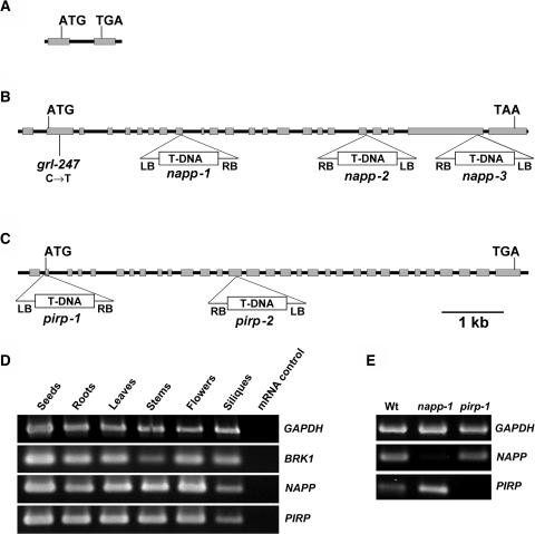
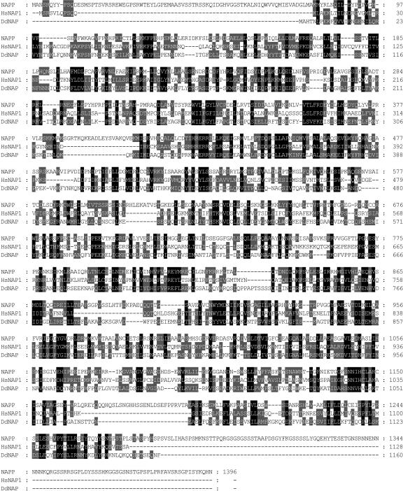
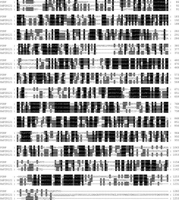
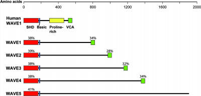
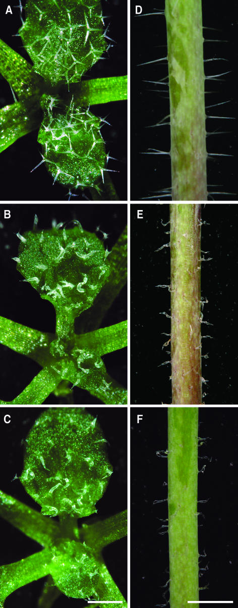
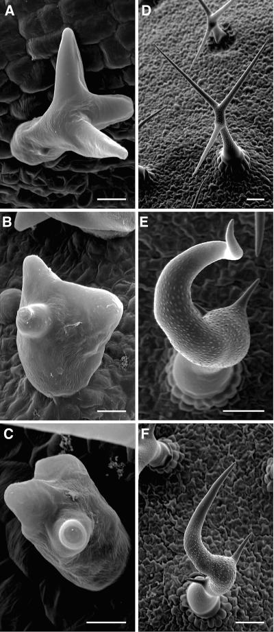
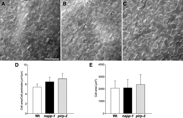
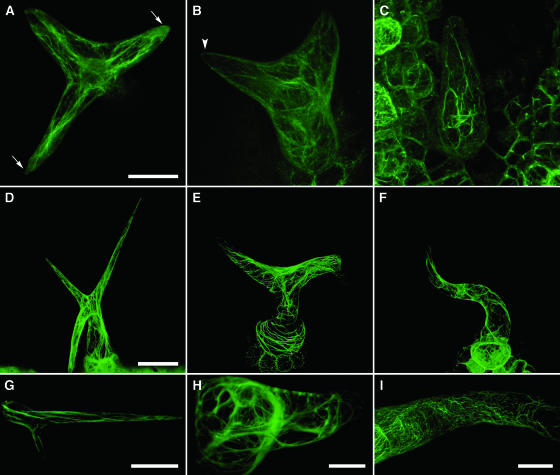
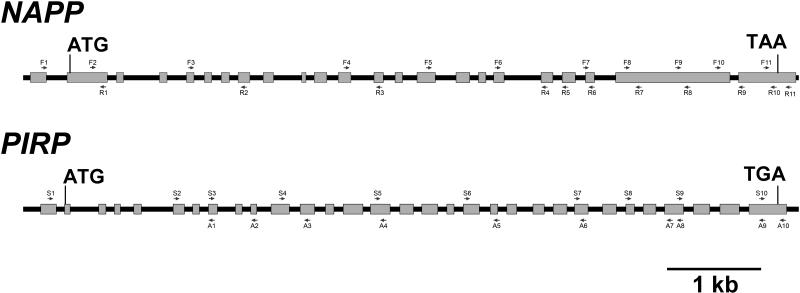
The ARP2/3 complex is an important regulator of actin nucleation and branching in eukaryotic organisms. All seven subunits of the ARP2/3 complex have been identified in Arabidopsis thaliana, and mutation of at least three of the subunits results in defects in epidermal cell expansion, including distorted trichomes. However, the mechanisms regulating the activity of the ARP2/3 complex in plants are largely unknown. In mammalian cells, WAVE and WASP proteins are involved in activation of the ARP2/3 complex. WAVE1 activity is regulated by a protein complex containing NAP1/HEM/KETTE/GEX-3 and PIR121/Sra-1/CYFIP/GEX-2. Here, we show that the WAVE1 regulatory protein complex is partly conserved in plants. We have identified Arabidopsis genes encoding homologs of NAP1 (NAPP), PIR121 (PIRP), and HSPC300 (BRK1). T-DNA inactivation of NAPP and PIRP results in distorted trichomes, similar to ARP2/3 complex mutants. The napp-1 mutant is allelic to the distorted mutant gnarled. The actin cytoskeleton in napp-1 and pirp-1 mutants shows orientation defects and increased bundling compared with wild-type plants. The results presented show that activity of the ARP2/3 complex in plants is regulated through an evolutionarily conserved mechanism.


Figures

Figure 1.
Figure 1.
Molecular Organization and Expression Analysis of BRK1, NAPP, and PIRP and Characterization of napp and pirp Mutants. (A), (B), and (C) Gene structure and location of mutations in BRK1 (A), NAPP (B), and PIRP (C). Gray boxes represent exons, and black lines represent introns and untranscribed flanking sequences. The location and orientation of T-DNA insertions in NAPP and PIRP are shown. grl-247 is an ethyl methanesulfonate–generated allele. (D) RT-PCR analysis of NAPP, PIRP, and BRK1 expression in various tissues. mRNA was extracted from different parts of Arabidopsis plants and used for RT-PCR analysis as described in the text. Glyceraldehyde-3-phosphate dehydrogenase (GAPDH) was used as a positive control. mRNA not subjected to reverse transcription was used as a negative control for each gene. (E) RT-PCR analysis of NAPP and PIRP expression in wild-type napp-1 and pirp-1. mRNA was extracted from leaves of soil-grown wild-type plants and homozygous mutant plants and subjected to RT-PCR. GAPDH was used as a positive control.
Figure 2.
Figure 2.
Amino Acid Alignment of NAPP. NAPP aligned with human Nap1 (HsNAP1) and Dictyostelium Nap (DdNAP). Amino acid residues in black indicate identity, and those in gray indicate conserved substitutions.
Figure 3.
Figure 3.
Amino Acid Alignment of PIRP. PIRP aligned with human PIR121 (HsPIR121) and Dictyostelium PIR121 (DdPIR121). Amino acid residues in black indicate identity, and those in gray indicate conserved substitutions.
Figure 4.
Figure 4.
Domain Structure of the Putative Arabidopsis WAVE Homologs. Comparison of the domain structure of human WAVE1 and the putative Arabidopsis WAVE homologs. The SHD domain, the basic region, the Pro-rich region, and the VCA domain are shown. The percentage of conserved amino acid residues between human WAVE1 and the putative Arabidopsis WAVE homologs are indicated for the SHD and the VCA domains. Arabidopsis ID numbers are as follows: WAVE1 (At2g34150), WAVE2 (At1g29170), WAVE3 (At5g01730), WAVE4 (At2g38440), and WAVE5 (At4g18600).
Figure 5.
Figure 5.
napp-1 and pirp-1 Plants Exhibit Defects in Trichome Development. (A) Trichomes on 2-week-old wild-type leaves have a uniform shape, generally with three branches. (B) and (C) Trichomes on 2-week-old leaves of napp-1 (B) and pirp-1 (C) have highly variable shapes, whereas leaf shape is normal. (D) Stem trichomes of wild-type plants are straight. (E) and (F) Stem trichomes of napp-1 (E) and pirp-1 (F) plants are shorter and crooked. Bars = 0.5 mm in (A), (B), and (C) and 1 mm in (D), (E), and (F).
Figure 6.
Figure 6.
Scanning Electron Microscopy Analyses of Wild-Type, napp-1, and pirp-1 Trichomes. (A) Developing wild-type trichome with elongating branches. (B) and (C) Developing napp-1 (B) and pirp-1 (C) trichomes (stage 4); branch initiation is apparently normal, but branch elongation is delayed/reduced, and trichome stalk diameter is increased. (D) Mature wild-type trichome with a thin stalk and branches of similar length. (E) and (F) Mature napp-1 (E) and pirp-1 (F) trichomes with increased stalk diameter, irregular branch position, and highly reduced secondary and tertiary branch lengths. Bars = 10 μm in (A), (B), and (C) and 50 μm in (D), (E), and (F).
Figure 7.
Figure 7.
Pavement Cell Lobe Structure in Wild-Type and napp/pirp Mutants. (A), (B), and (C) Leaf pavement cell shapes of wild-type (A), napp-1 (B), and pirp-2 (C) plants. The fourth rosette leaf of 2-week-old plants was cleared, and the adaxial pavements cells were observed using phase-contrast light microscopy. Bar = 50 μm. (D) Comparison of lobe extensions in wild-type and mutant leaf pavement cells. The area and the perimeter were measured on 118 pavement cells in wild-type and mutant plants, and the ratio between area and perimeter was calculated for each cell. (E) Comparison of cell area of wild-type and mutant pavement cells.
Figure 8.
Figure 8.
Organization of F-Actin in Leaf Trichomes of Wild-Type, napp-1, and pirp-1 Plants. F-actin was visualized by stable expression of YFP-mTalin in 8-d-old plants ([A] to [C]) and 9-d-old plants ([D] to [F]) and by transient expression in 14-d-old plants ([G] to [I]). Young ([A] to [C]) and mature ([D] to [I]) trichomes were imaged using confocal microscopy. Three-dimensional projections were generated from multiple optical sections. (A) A young wild-type trichome with elongating branches. Actin filaments are longitudinally oriented; two of the branch tips are dominated by diffuse actin. (B) A pirp-1 trichome at a similar stage of development as in (A). Actin filaments show a more random orientation but not increased bundling. Diffuse actin is not observed in the branch tips. (C) A pirp-1 trichome at a slightly later stage contains thick actin cables extending both longitudinally and transversely across the branch. (D) A (mature) wild-type trichome showing a fine network of longitudinally oriented actin filaments. (E) A pirp-1 trichome with thick actin cables that are mainly transversely oriented, especially in the radially expanded stalk. Note the high density of actin filaments at the branch point between the two branches. (F) A napp-1 trichome showing transversely oriented filaments. (G) A branch of a wild-type trichome with a population of diffuse actin at the branch tip. (H) A transverse cross section through a pirp-1 trichome. Thick actin cables extend across the stalk. The branch tip has small amounts of both actin filaments and diffuse actin. (I) Portion of the branch of a moderately affected napp-1 trichome. Thick actin cables are not seen, but actin filaments show varying degrees of transverse orientation. Bars = 20 μm in (A) to (C), (H), and (I), 50 μm in (D) to (F), and 40 μm in (G).
Figure 9.
Figure 9.
Position of NAPP and PIRP Primers Used in This Study. The arrows indicate the orientation of the primers.

References

    1. Alonso, J.M., et al. (2003). Genome-wide insertional mutagenesis of Arabidopsis thaliana. Science 301, 653–657. - PubMed
    1. Amann, K.J., and Pollard, T.D. (2001). The Arp2/3 complex nucleates actin filament branches from the sides of pre-existing filaments. Nat. Cell Biol. 3, 306–310. - PubMed
    1. Baumgartner, S., Martin, D., Chiquet-Ehrismann, R., Sutton, J., Desai, A., Huang, I., Kato, K., and Hromas, R. (1995). The HEM proteins: A novel family of tissue-specific transmembrane proteins expressed from invertebrates through mammals with an essential function in oogenesis. J. Mol. Biol. 251, 41–49. - PubMed
    1. Bechtold, N., Ellis, J., and Pelletier, G. (1993). In planta Agrobacterium mediated gene transfer by infiltration of adult Arabidopsis thaliana plants. CR Acad. Sci. III–Vie 316, 1194–1199.
    1. Blagg, S.L., Stewart, M., Sambles, C., and Insall, R.H. (2003). PIR121 regulates pseudopod dynamics and SCAR activity in Dictyostelium. Curr. Biol. 13, 1480–1487. - PubMed

Publication types

MeSH terms