. 2001 Nov;67(11):4984-91.
doi: 10.1128/AEM.67.11.4984-4991.2001.

Identification by subtractive hybridization of sequences specific for Salmonella enterica serovar enteritidis

Affiliations

Identification by subtractive hybridization of sequences specific for Salmonella enterica serovar enteritidis

P G Agron et al. Appl Environ Microbiol. 2001 Nov.

Abstract

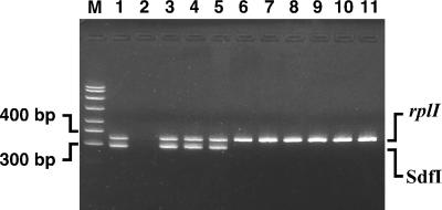
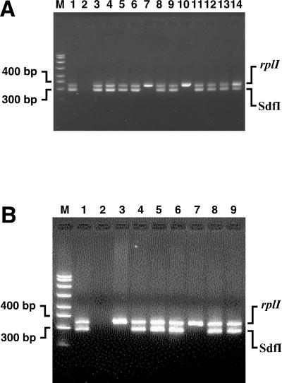
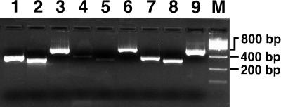
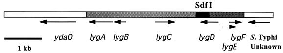
Salmonella enterica serovar Enteritidis, a major cause of food poisoning, can be transmitted to humans through intact chicken eggs when the contents have not been thoroughly cooked. Infection in chickens is asymptomatic; therefore, simple, sensitive, and specific detection methods are crucial for efforts to limit human exposure. Suppression subtractive hybridization was used to isolate DNA restriction fragments present in Salmonella serovar Enteritidis but absent in other bacteria found in poultry environments. Oligonucleotide primers to candidate regions were used in polymerase chain reactions to test 73 non-Enteritidis S. enterica isolates comprising 34 different serovars, including Dublin and Pullorum, two very close relatives of Enteritidis. A primer pair to one Salmonella difference fragment (termed Sdf I) clearly distinguished serovar Enteritidis from all other serovars tested, while two other primer pairs only identified a few non-Enteritidis strains. These primer pairs were also useful for the detection of a diverse collection of clinical and environmental Salmonella serovar Enteritidis isolates. In addition, five bacterial genera commonly found with Salmonella serovar Enteritidis were not detected. By treating total DNA with an exonuclease that degrades sheared chromosomal DNA but not intact circular plasmid DNA, it was shown that Sdf I is located on the chromosome. The Sdf I primers were used to screen a Salmonella serovar Enteritidis genomic library and a unique 4,060-bp region was defined. These results provide a basis for developing a rapid, sensitive, and highly specific detection system for Salmonella serovar Enteritidis and provide sequence information that may be relevant to the unique characteristics of this serovar.


Figures

FIG. 1
FIG. 1
Specificity of S. enterica serovar Enteritidis detection determined using the Sdf I primer pair in PCRs. Lane M, size standards; lane 1, Salmonella serovar Enteritidis CAHFS-546 (phage type 8); lane 2, no template; lane 3, Salmonella serovar Enteritidis CAHFS-184 (phage type 4); lane 4, Salmonella serovar Enteritidis 97-6371A (phage type 8); lane 5, Salmonella serovar Enteritidis 97-1866IN (phage type 13A); lane 6, Salmonella serovar Pullorum; lane 7, Salmonella serovar Typhimurium; lane 8, Salmonella serovar Heidelberg; lane 9, Salmonella serovar Montevideo; lane 10, E. coli; lane 11, C. freundii. Amplicons produced by the Sdf I primers (293 bp) and the rplI primers (343 bp) are indicated.
FIG. 2
FIG. 2
Specificity of detection of selected Salmonella serovar Enteritidis phage type reference strains determined using the Sdf I primer pair in PCRs. The strains used are from the NVSL unless indicated by a specific designation. (A) Detection of Sdf I in phage type reference strains. Lane M, size markers; lane 1, CAHFS-546 (positive control); lane 2, no template; lane 3, phage type 2; lane 4, phage type 3; lane 5, phage type 4; lane 6, phage type 6; lane 7, phage type 6A; lane 8, phage type 8; lane 9, phage type 9; lane 10, phage type 9A; lane 11, phage type 13A; lane 12, 95-13141 (phage type 14B); lane 13, phage type 24; lane 14, phage type 34. (B) Detection of Sdf I in phage type reference strains and clinical strains of phage types 6A, 6B, and 9A. Lane M, size markers; lane 1, CAHFS-546 (positive control); lane 2, no template; lane 3, NVSL 9 (phage type 6A); lane 4, CAHFS-435 (phage type 6A); lane 5, CAHFS-436 (phage type 6A); lane 6, CAHFS-739 (phage type 6B); lane 7, NVSL 13 (phage type 9A); lane 8, D0144-CDC (phage type 9A); lane 9, D01760-CDC (phage type 9A). Amplicons produced by the Sdf I primers (293 bp) and rplI primers (343 bp) are indicated.
FIG. 3
FIG. 3
The Salmonella serovar Enteritidis Sdf I region is located on the chromosome. Lane 1, total DNA amplified with rplI primers (343-bp amplicon); lane 2, total DNA amplified with the Sdf I primers (293-bp amplicon); lane 3, total DNA amplified with the spvC primers (565-bp amplicon); lane 4, plasmid preparation treated with exo-DNase amplified with rplI primers; lane 5, plasmid preparation treated with exo-DNase and amplified with Sdf I primers; lane 6, plasmid preparation treated with exo-DNase and amplified with spvC primers. Lanes 7, 8, and 9 are the same as lanes 4, 5, and 6, respectively, but without exo-DNase treatment before amplification. Strain CAHFS-285 (phage type 4) was used for these experiments.
FIG. 4
FIG. 4
Chromosomal context of Sdf I. Schematic representation of the Sdf I region from Salmonella serovar Enteritidis CAHFS-285 (phage type 4). Open boxes indicate sequence with identity to S. enterica serovar Typhi. Gray and black boxes indicate novel sequences. Sdf I, bounded by Sau3AI sites, is shown in black. All ORFs of more than 100 codons are indicated with black arrows.

References

    1. Akopyants N S, Fradkov A, Diatchenko L, Hill J E, Siebert P D, Lukyanov S A, Sverdlov E D, Berg D E. PCR-based subtractive hybridization and differences in gene content among strains of Helicobacter pylori. Proc Natl Acad Sci USA. 1998;95:13108–13113. - PMC - PubMed
    1. Bäumler A J, Hargis B M, Tsolis R M. Tracing the origins of Salmonella outbreaks. Science. 2000;287:50–52. - PubMed
    1. Belgrader P, Benett W, Hadley D, Richards J, Stratton P, Mariella R, Jr, Milanovich F. PCR detection of bacteria in seven minutes. Science. 1999;284:449–450. - PubMed
    1. Birnboim H C, Doly J. A rapid alkaline extraction procedure for screening recombinant plasmid DNA. Nucleic Acids Res. 1979;7:1513–1523. - PMC - PubMed
    1. Dibb-Fuller M P, Allen-Vercoe E, Thorns C J, Woodward M J. Fimbriae- and flagella-mediated association with and invasion of cultured epithelial cells by Salmonella enteritidis. Microbiology. 1999;145:1023–1031. - PubMed

Publication types

MeSH terms

Associated data

LinkOut - more resources