. 2001 Jul 31;98(16):8966-71.
doi: 10.1073/pnas.151105198. Epub 2001 Jul 17.

Trace amines: identification of a family of mammalian G protein-coupled receptors

Affiliations

Trace amines: identification of a family of mammalian G protein-coupled receptors

B Borowsky et al. Proc Natl Acad Sci U S A. .

Abstract

Tyramine, beta-phenylethylamine, tryptamine, and octopamine are biogenic amines present in trace levels in mammalian nervous systems. Although some "trace amines" have clearly defined roles as neurotransmitters in invertebrates, the extent to which they function as true neurotransmitters in vertebrates has remained speculative. Using a degenerate PCR approach, we have identified 15 G protein-coupled receptors (GPCR) from human and rodent tissues. Together with the orphan receptor PNR, these receptors form a subfamily of rhodopsin GPCRs distinct from, but related to the classical biogenic amine receptors. We have demonstrated that two of these receptors bind and/or are activated by trace amines. The cloning of mammalian GPCRs for trace amines supports a role for trace amines as neurotransmitters in vertebrates. Three of the four human receptors from this family are present in the amygdala, possibly linking trace amine receptors to affective disorders. The identification of this family of receptors should rekindle the investigation of the roles of trace amines in mammalian nervous systems and may potentially lead to the development of novel therapeutics for a variety of indications.


Figures

Figure 1
Figure 1
Alignment of rat, mouse, and human TA1 and rat TA2 receptors (GenBank accession nos. AF380186, AF380187, AF380185, and AF380188, respectively). Shaded residues are conserved in all four receptors. Triangles and circles indicate residues conserved in TA1–TA15. Open triangles are also conserved among all human monaminergic receptors, and open circles are conserved among all human 5-HT but not NE or DA receptors. Seven putative TM domains are indicated.
Figure 2
Figure 2
Voltage-clamp responses to trace amines in oocytes. (A) Response to 100 μM octopamine (Oct) or 5-HT in an oocyte expressing rat TA1 and CFTR. (B) Response to 100 nM tyramine (Tyr) in an oocyte expressing rat TA1 and CFTR. (C) Response to 100 μM octopamine in an oocyte expressing only TA1. (D) Response to 100 nM tyramine (Tyr) in an oocyte expressing human TA1 and CFTR. Holding potential was −80 mV for all oocytes. Marker bar in D also applies to A and C.
Figure 3
Figure 3
β-PEA-induced responses in COS-7 cells transfected with human TA1, rat TA2, or vector. Cells were incubated with increasing concentrations of β-PEA and cAMP accumulation measured. Data are from duplicate determinations and are representative of three to six experiments.
Figure 4
Figure 4
A phylogenetic tree for trace amine receptors TA1–TA15, human 5-HT receptors 5-HT1a, 5-HT1b, 5-HT1d, 5-HT4, 5-HT5, 5-HT7, human α1a receptor (AR-α1a), GPR57, GPR58, PNR, 5-HT4 psuedogene (5-HT), drosophila (Drosoph) receptors for octopamine (Oct), 5-HT1 and tyramine (Tyr), Caenorhabditis elegans (C. Elegans) 5-HT receptor, tyramine receptors from bee (Bee Tyr) and locust (Locust Tyr), and a snail octopamine receptor (Snail Oct). Amino acid sequences for each receptor spanning from the start of TMI to the end of TMVII were aligned by using the CLUSTALW algorithm and the tree constructed by the NJ method on a DecypherII Bioaccelerator (TimeLogic, Reno, NV). GenBank accession nos.: AF380190 (rat TA3), AF380189 (human TA3), AF380191 (rat TA4), AF380192 (human TA4), AF380193-AF380203 (TA5–TA15).
Figure 5
Figure 5
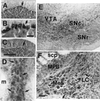
Photomicrographs showing hybridization signals for TA1 mRNA in mouse CNS. Signal detected in cerebellar Purkinje cells (arrows) hybridized with antisense (A and B) and sense (C) probes. Scale bar in C (200 μm) also applies to A and E; scale bar in B, 25 μm. Photomicrographs showing hybridization signal in cells (arrows) in the dorsal raphe (D), ventral tegmental area (VTA), substantia nigra, compact part (SNc) and reticular part (SNr) (E), and locus coeruleus (LC; F). Scale bar in F (50 μm) also applies to D. MPB, medial parabrachial nucleus; Me5, mesencephalic trigeminal nucleus; m, medial longitudinal fasciculus; scp, superior cerebral peduncle.

References

    1. Usdin E, Sandler M, editors. Trace Amines and the Brain. New York: Dekker; 1976.
    1. Boulton A A. In: Trace Amines and the Brain. Usdin E, Sandler M, editors. New York: Dekker; 1976.
    1. Juorio A V. Brain Res. 1976;111:442–445. - PubMed
    1. Durden D A, Philips S R. J Neurochem. 1980;34:1725–1732. - PubMed
    1. Grimsby J, Toth M, Chen K, Kumazawa T, Klaidman L, Adams J D, Karoum F, Gal J, Shih J C. Nat Genet. 1997;17:206–210. - PubMed