. 2002 Feb;109(4):533-40.
doi: 10.1172/JCI14099.

Cystin, a novel cilia-associated protein, is disrupted in the cpk mouse model of polycystic kidney disease

Affiliations

Cystin, a novel cilia-associated protein, is disrupted in the cpk mouse model of polycystic kidney disease

Xiaoying Hou et al. J Clin Invest. 2002 Feb.

Abstract

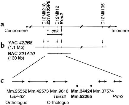
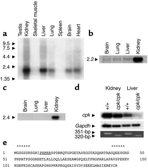
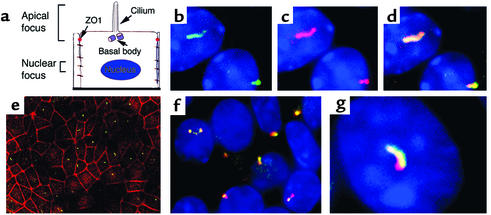
The congenital polycystic kidney (cpk) mutation is the most extensively characterized mouse model of polycystic kidney disease (PKD). The renal cystic disease is fully expressed in homozygotes and is strikingly similar to human autosomal recessive PKD (ARPKD), whereas genetic background modulates the penetrance of the corresponding defect in the developing biliary tree. We now describe the positional cloning, mutation analysis, and expression of a novel gene that is disrupted in cpk mice. The cpk gene is expressed primarily in the kidney and liver and encodes a hydrophilic, 145-amino acid protein, which we term cystin. When expressed exogenously in polarized renal epithelial cells, cystin is detected in cilia, and its expression overlaps with polaris, another PKD-related protein. We therefore propose that the single epithelial cilium is important in the functional differentiation of polarized epithelia and that ciliary dysfunction underlies the PKD phenotype in cpk mice.


Figures

Figure 1
Figure 1
Integrated genetic and physical map of the cpk candidate interval. Recombination analyses refined the cpk locus to an approximately 100-kb interval, centered on D12Mit12 and delimited by 221A10SP6 and Rrm2 (indicated in bold italics; a). These data positioned cpk on a single BAC, 221A10, within our yeast and bacterial artificial cromosome (YAC/BAC)-based physical map (b). Computational analyses identified six putative transcriptional units within the cpk interval, and each predicted gene corresponded, at least in part, to a mouse UniGene cluster (c).
Figure 2
Figure 2
Organization of the cpk genomic sequence and identification of the cpk mutation. (a) Alignment of the cpk cDNA, the UniGene consensus sequences, and the BAC genomic sequence demonstrated that the cpk gene is encoded in five exons spanning 14.4 kb of genomic DNA. The first nucleotide of the cDNA corresponds to the first nucleotide of exon 1, which spans 1,184 bp and is the largest of the five exons. This exon contains an ATG start site that lies within a Kozak consensus sequence (CGCGCCatgG). The 435-bp ORF extends into exon 3. Exons 4 and 5 are apparently untranslated, and a putative cryptic splice site (gaacagCTG) within exon 5 appears to account for the 1,856-bp and 1,786-bp (gray box) splice variants. An atypical polyadenylation signal (ATTAAA) lies 22-nt upstream of the poly(A+) tail. Of note, the microsatellite marker, D12Mit12, lies within intron 1 of the cpk gene. (b) PCR amplification and direct sequence analysis identified tandem 12-bp and 19-bp deletions in exon 1 of the cpk gene. The comparative sequence is indicated in bold text. The resulting frameshift truncates the predicted protein. The position of the PCR primers is identified by arrows. The putative Kozak sequence is underlined and italicized. (c) Primers flanking the tandem deletion in the cpk mutant allele amplify a 351-bp product from wild-type (B6 and D2) DNA, a 320-bp product from B6-cpk/cpk DNA, and both bands from B6-+/cpk and F1 +/cpk heterozygotes. In the key F2 cpk/cpk recombinants (R1–R5), only the 320-bp mutant allele was amplified.
Figure 3
Figure 3
Expression pattern of the cpk transcript and characterization of the predicted protein product. (a) The tissue distribution of the 2.2-kb cpk transcript is shown in adult mouse tissues. The size of the polyadenylated transcript is consistent with the 1,856-bp full-length cDNA. The size markers are indicated on the left and expressed as kilobases. (b) Northern blot analysis of mouse fetal poly(A+) RNA revealed a 2.2-kb transcript in the fetal kidney. (c) Northern blot analysis of human fetal poly(A+) RNA revealed a 2.4-kb transcript in the fetal kidney. (d) The relative expression of the 2.2-kb cpk transcript is shown in kidney and liver poly(A+) RNA from 2-week-old B6-WT (n = 5 mice) and B6-cpk/cpk (n = 5 mice) (top panel). Rehybridization with Gapdh cDNA served as a loading control (middle panel). RT-PCR was performed using the deletion-flanking primer pairs on the same poly(A+) RNAs (bottom panel). (e) Amino acid sequence of the predicted 145-AA protein product. Two potential myristoylation sites (residues 2–7, 43–48, indicated by asterisks) are predicted, the first of which is coupled to a polybasic domain (underlined).
Figure 4
Figure 4
Localization of exogenously expressed cystin in stably transfected mCCD cells. To analyze cystin localization, wild-type mCCD cells were transfected with a myc and his epitope–tagged cystin construct. Stable cell cultures were established by selection in Blasticidin. Immunofluorescence analysis was conducted in cells grown on cell culture inserts for a minimum of 3 days after confluence to allow cilial development. (a) Schematic diagram of a principal cell with its primary apical cilium. The two focal planes used in immunofluorescence imaging are indicated. The apical focal plane was used to capture the cilium and the position of the tight junction, indicated by α-ZO-1 staining, and the nuclear focal plane identified the HOECHST-stained nuclei (blue). (b) Immunofluorescence localization of cystin (green) as determined using the anti-his rabbit polyclonal Ab. (c) Cystin (red) localization in the same mCCD cells as shown in b when probed with the anti-myc mAb. (d). Merged image of b and c demonstrated colocalization (yellow) of the myc and his epitope–tagged cystin. (e) In a broad-field view, staining with the anti-his polyclonal Ab (green) indicated that cystin localized to the center of mCCD cells relative to the tight junctions stained for ZO-1 (red). (f and g) Broad-field and representative high-magnification views demonstrated the colocalization of the exogenously expressed cystin (red, anti-myc mAb) and endogenous polaris (green, rabbit polyclonal Ab) in cilia of mCCD cells.

References

    1. Gabow P. Autosomal polycystic kidney disease. N Engl J Med. 1993;329:332–342. - PubMed
    1. Watnick T, Germino G. Molecular basis of autosomal dominant polycystic kidney disease. Semin Nephrol. 1999;19:327–343. - PubMed
    1. Guay-Woodford, L., Jafri, Z., and Bernstein, J. 1999. Other cystic diseases. In Comprehensive clinical nephrology. R. Johnson and J. Feehally, editors. Mosby International. London, United Kingdom. 50.1–50.2.
    1. Zerres K, et al. Mapping of the gene for autosomal recessive polycystic kidney disease (ARPKD) to chromosome 6p21-cen. Nat Genet. 1994;7:429–432. - PubMed
    1. Guay-Woodford L, et al. The severe perinatal form of autosomal recessive polycystic kidney disease (ARPKD) maps to chromosome 6p21.1-p12: implications for genetic counseling. Am J Hum Genet. 1995;56:1101–1107. - PMC - PubMed

Publication types

MeSH terms

Associated data