. 2001 Dec;11(12):2050-8.
doi: 10.1101/gr.194701.

Genomic characterization of recent human LINE-1 insertions: evidence supporting random insertion

Affiliations

Genomic characterization of recent human LINE-1 insertions: evidence supporting random insertion

I Ovchinnikov et al. Genome Res. 2001 Dec.

Abstract

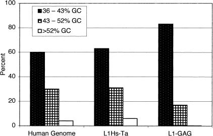
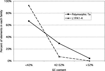
LINE-1 (L1) elements play an important creative role in genomic evolution by distributing both L1 and non-L1 DNA in a process called retrotransposition. A large percentage of the human genome consists of DNA that has been dispersed by the L1 transposition machinery. L1 elements are not randomly distributed in genomic DNA but are concentrated in regions with lower GC content. In an effort to understand the consequences of L1 insertions, we have begun an investigation of their genomic characteristics and the changes that occur to them over time. We compare human L1 insertions that were created either during recent human evolution or during the primate radiation. We report that L1 insertions are an important source for the creation of new microsatellites. We provide evidence that L1 first strand cDNA synthesis can occur from an internal priming event. We note that in contrast to older L1 insertions, recent L1s are distributed randomly in genomic DNA, and the shift in the L1 genomic distribution occurs relatively rapidly. Taken together, our data indicate that strong forces act on newly inserted L1 retrotransposons to alter their structure and distribution.


Figures

Figure 1
Figure 1
L1 display, method, and results. (A) L1 display. A L1Hs-Ta insertion (rectangle) is depicted surrounded by flanking DNA (solid lines). The broken lines represent the products of two rounds of PCR amplifications. The arrows below indicate the relative positions and orientations of the primers. (B) L1 display results. A typical L1 display experiment performed with a single decamer on genomic DNA from 42 individuals is shown. One fixed (solid line) and two polymorphic (broken lines) L1Hs-Ta insertions can be seen.
Figure 1
Figure 1
L1 display, method, and results. (A) L1 display. A L1Hs-Ta insertion (rectangle) is depicted surrounded by flanking DNA (solid lines). The broken lines represent the products of two rounds of PCR amplifications. The arrows below indicate the relative positions and orientations of the primers. (B) L1 display results. A typical L1 display experiment performed with a single decamer on genomic DNA from 42 individuals is shown. One fixed (solid line) and two polymorphic (broken lines) L1Hs-Ta insertions can be seen.
Figure 2
Figure 2
Organization of the L1 insertion H15/B10-800. The L1 display clone (H15/B10-800) contains the 3′ end of a L1Hs-Ta followed by a 23-bp poly(A) tail and 616-bp of 3′ flanking DNA. GenBank accession no. AC027480 contains a full-length L1Hs-Ta element followed by a 23-bp long poly(A) tail and the proximal 211 bp of 3′-flanking DNA from the H15/B10-800 L1 insertion. More distal 3′ flanking DNA is not homologous to the distal 3′ flanking DNA from H15/B10-800. GenBank accession no. AC026092 is the locus where H15/B10-800 inserted. It contains only the distal 405 bp of the H15/B10-800 3′ flanking DNA. The horizontal arrows indicate the positions of the PCR primers that were used to confirm the structure of the H15/B10-800 integration. The A-rich sequences present at each of the loci between the proximal and distal 3′-flanking DNA are indicated.
Figure 3
Figure 3
Presence of various types of DNA repeat sequences in the 3′-flanking DNA of L1 insertions. The presence of repeat sequences in the 3′-flanking DNA of L1 insertions was determined by the RepeatMasker program. L1Hs-Ta insertions are represented in the black bars; L1-GAG insertions are represented in the stippled bars.
Figure 4
Figure 4
Distance from the poly(A) addition signal to the nearest repeat sequence in 3′-flanking DNA. The distance was calculated in bp from the end of the poly(A) addition signal. L1Hs-Ta insertions are represented in the black bars; L1-GAG insertions are represented in the stippled bars.
Figure 5
Figure 5
Distribution of L1 insertions in genomic DNA of different GC content. The GC content of the 3′ flanks of L1 insertions was calculated starting 150 bp after the poly(A) addition signal. Each insertion was assigned to bins of either 36%–43% GC (stippled), 43%–52% GC (checkered), or >52% GC (open box).
Figure 6
Figure 6
Comparison of the distribution of younger and older L1 insertions in genomic DNA of different GC content. The GC content of 10 kb surrounding the insertion sites (5 kb of both the 5′ and 3′ flanks) of polymorphic L1Hs–Ta and GAG elements was calculated. Each insertion was assigned to different bins of GC content. For this analysis we analyzed 24 polymorphic insertions, and 57 GAG insertions; the available flanking sequences of one GAG and five polymorphic elements were too short to be included.

References

    1. Adams JW, Kaufman RE, Kretschmer PJ, Harrison M, Nienhuis AW. A family of long reiterated DNA sequences, one copy of which is next to the human beta globin gene. Nucleic Acids Res. 1980;8:6113–6128. - PMC - PubMed
    1. Altschul SF, Gish W, Miller W, Myers EW, Lipman DJ. Basic local alignment search tool. J Mol Biol. 1990;215:403–410. - PubMed
    1. Arcot SS, Adamson AW, Risch GW, LaFleur J, Robichaux MB, Lamerdin JE, Carrano AV, Batzer MA. High-resolution cartography of recently integrated human chromosome 19—specific Alu fossils. J Mol Biol. 1998;281:843–856. - PubMed
    1. Arcot SS, Wang Z, Weber JL, Deininger PL, Batzer MA. Alu repeats: A source for the genesis of primate microsatellites. Genomics. 1995;29:136–144. - PubMed
    1. Bailey JA, Carrel L, Chakravarti A, Eichler EE. Molecular evidence for a relationship between LINE-1 elements and X chromosome inactivation: The Lyon repeat hypothesis. Proc Natl Acad Sci. 2000;97:6634–6639. - PMC - PubMed

MeSH terms

Substances

Associated data