

Bacterial community structure and location in Stilton cheese
- PMID: 12788761
- PMCID: PMC161494
- DOI: 10.1128/AEM.69.6.3540-3548.2003
Bacterial community structure and location in Stilton cheese
Abstract
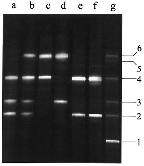
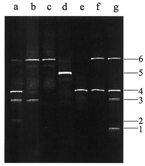
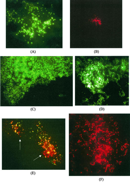
The microbial diversity occurring in Stilton cheese was evaluated by 16S ribosomal DNA analysis with PCR-denaturing gradient gel electrophoresis. DNA templates for PCR experiments were directly extracted from the cheese as well as bulk cells harvested from a variety of viable-count media. The variable V3 and V4-V5 regions of the 16S genes were analyzed. Closest relatives of Lactococcus lactis, Enterococcus faecalis, Lactobacillus plantarum, Lactobacillus curvatus, Leuconostoc mesenteroides, Staphylococcus equorum, and Staphylococcus sp. were identified by sequencing of the DGGE fragments. Fluorescently labeled oligonucleotide probes were developed to detect Lactococcus lactis, Lactobacillus plantarum, and Leuconostoc mesenteroides in fluorescence in situ hybridization (FISH) experiments, and their specificity for the species occurring in the community of Stilton cheese was checked in FISH experiments carried out with reference cultures. The combined use of these probes and the bacterial probe Eub338 in FISH experiments on Stilton cheese sections allowed the assessment of the spatial distribution of the different microbial species in the dairy matrix. Microbial colonies of bacteria showed a differential location in the different parts of the cheese examined: the core, the veins, and the crust. Lactococci were found in the internal part of the veins as mixed colonies and as single colonies within the core. Lactobacillus plantarum was detected only underneath the surface, while Leuconostoc microcolonies were homogeneously distributed in all parts observed. The combined molecular approach is shown to be useful to simultaneously describe the structure and location of the bacterial flora in cheese. The differential distribution of species found suggests specific ecological reasons for the establishment of sites of actual microbial growth in the cheese, with implications of significance in understanding the ecology of food systems and with the aim of achieving optimization of the fermentation technologies as well as preservation of traditional products.
Figures
References
-
- Albenzio, M., M. R. Corbo, S. U. Rehman, P. F. Fox, M. De Angelis, A. Corsetti, A. Sevi, and M. Gobetti. 2001. Microbiological and biochemical characteristics of Canestrato Pugliese cheese made from raw milk, pasteurized milk or by heating the curd in hot whey. Int. J. Food Microbiol. 67:35-48. - PubMed
-
- Amann, R., B. M. Fuchs, and S. Behrens. 2001. The identification of microorganisms by fluorescence in situ hybridisation. Curr. Opin. Biotechnol. 12:231-236. - PubMed
Publication types
MeSH terms
Substances
Associated data
- Actions
- Actions
- Actions
- Actions
- Actions
- Actions
- Actions
- Actions
- Actions
- Actions
- Actions
LinkOut - more resources
Full Text Sources
Other Literature Sources
Molecular Biology Databases
