. 2006 Oct;18(10):2469-78.
doi: 10.1105/tpc.106.045906. Epub 2006 Oct 13.

A rice Tc1/mariner-like element transposes in yeast

Affiliations

A rice Tc1/mariner-like element transposes in yeast

Guojun Yang et al. Plant Cell. 2006 Oct.

Abstract

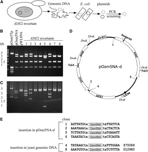
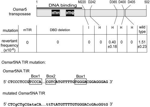
The Tc1/mariner transposable element superfamily is widely distributed in animal and plant genomes. However, no active plant element has been previously identified. Nearly identical copies of a rice (Oryza sativa) Tc1/mariner element called Osmar5 in the genome suggested potential activity. Previous studies revealed that Osmar5 encoded a protein that bound specifically to its own ends. In this report, we show that Osmar5 is an active transposable element by demonstrating that expression of its coding sequence in yeast promotes the excision of a nonautonomous Osmar5 element located in a reporter construct. Element excision produces transposon footprints, whereas element reinsertion occurs at TA dinucleotides that were either tightly linked or unlinked to the excision site. Several site-directed mutations in the transposase abolished activity, whereas mutations in the transposase binding site prevented transposition of the nonautonomous element from the reporter construct. This report of an active plant Tc1/mariner in yeast will provide a foundation for future comparative analyses of animal and plant elements in addition to making a new wide host range transposable element available for plant gene tagging.


Figures

Figure 1.
Figure 1.
Scheme of Osmar5, the Osmar5 Transposase Coding Sequence (Osmar5 Transposase), and the Nonautonomous Element (Osmar5NA). TIRs are shown as black triangles. White boxes represent Osmar5 coding exons, and shaded regions represent noncoding sequences; slashed regions indicate introns. The dark region in Osmar5NA represents the linker sequence (see Methods). The NdeI site used for ADE2 revertant plasmid digestion is also shown. HTH1 and HTH2 represent helix-turn-helix motifs 1 and 2, respectively. Box1 and Box2 indicate transposase binding site motifs. The three Asp residues (D242, D365, and D405) constitute the putative DD39D motif.
Figure 2.
Figure 2.
Yeast Transposition Assay Constructs and Protocol. The positions of primers used for PCR analysis in Figure 3B are shown as gray arrows. amp, ampicillin resistance gene; ARS1, autonomous replication sequence1; ARS H4, autonomous replication sequence of the H4 gene; CEN6 and CEN4, centromere sequences of yeast chromosomes 6 and 4, respectively; cyc1 ter, terminator of yeast cyclin gene cyc1; OriEC, E. coli replication origin; Pgal1, yeast gal1 promoter; pRS413, control vector like pOsm5Tp but without the transposase. See Methods and text for details.
Figure 3.
Figure 3.
Osmar5NA Footprints. (A) ADE2 revertants on medium lacking adenine. The left two sectors show single colonies derived from two independent pOsm5Tp and pOsm5NA double transformant colonies. Sectors at right are from pRS413 and pOsm5NA double transformant colonies. (B) Agarose gel of PCR products from the ade2 5′ UTR of the ADE2 revertant plasmids. Expected band size is 1.4 or 0.4 kb (control), with or without Osmar5NA, respectively. (C) Sequences of excision sites of ADE2 revertants. Part of the sequence of pOsm5NA before excision is shown at top, including the ends of Osmar5NA (boxed) and flanking sequence. The dinucleotides TA that flank Osmar5 in the donor vector and in each footprint are shown in red.
Figure 4.
Figure 4.
Genomic DNA Gel Blot Analysis of ADE2 Revertants. Genomic DNA (from eight independent revertants, labeled 1 to 8) was digested with DraI, and blots were probed with Osmar5NA. Controls are untransformed yeast (DG2523) and pOsm5NA. Two minor bands in the vector control and revertant lanes are attributable to nonspecific cleavage by DraI. DNA size markers at left are in kilobases.
Figure 5.
Figure 5.
Reintegration Sites of Osmar5NA. (A) Scheme of plasmid rescue from ADE2 revertant genomic DNA. Yeast genomic DNA was extracted from ADE2 revertants and used to transform E. coli (see text for details). The small gray and black circles represent pOsm5NA and pOsm5Tp, respectively. (B) Agarose gel analysis of DraI digestion of the recovered pOsm5NA derivative plasmids from (A). DNA size markers are shown at left. (C) NdeI digestion of the plasmids used for (B). (D) Insertion sites in pOsm5NA derivatives (pOsm5NA-d); pWL89A lacks Osmar5NA. Note that Osmar5NA has a NdeI site but not a DraI site. (E) Insertion sites of ADE2 revertants in either the plasmid vector or yeast genomic DNA. Accession numbers of yeast genomic DNA are shown at right.
Figure 6.
Figure 6.
Mutations Introduced in the Transposase and TIR and Their Effect on Transposition. Vectors containing the intact Osmar5 transposase gene (wild type) and its mutated forms were cotransformed with pOsm5NA, and double transformants were selected for ADE2 reversion. mTIR, mutated TIR of Osmar5NA; DBD deletion, DNA binding domain deletion; M220→I, Met at position 220 mutated to Ile; D242→H, Asp at position 242 mutated to His; D365→H, Asp at position 365 mutated to His; D400→H, Asp at position 400 mutated to His; D405→H, Asp at position 405 mutated to His. Standard errors for six independent events are shown. The nucleotide changes in the Osmar5NA TIRs are shown in lowercase letters. Dots represent omitted internal sequences of Osmar5NA. Previously identified DNA binding motifs are shown in boxes.
Figure 7.
Figure 7.
Representative Footprints for Tc3, Mos1, Minos, and Osmar5NA. Donor sites are shown as double stranded, but footprints are shown as single strands (top strand). TSDs are shown in red. Lowercase letters indicate residues retained from transposon ends. Arrows indicate proven (Tc3 and Mos1) or predicted (Minos and Osmar5NA) excision cleavage sites. Based on Bryan et al. (1990), van Luenen et al. (1994), Arca et al. (1997), and Zagoraiou et al. (2001).

References

    1. Arca, B., Zabalou, S., Loukeris, T.G., and Savakis, C. (1997). Mobilization of a Minos transposon in Drosophila melanogaster chromosomes and chromatid repair by heteroduplex formation. Genetics 145 267–279. - PMC - PubMed
    1. Auge-Gouillou, C., Hamelin, M.H., Demattei, M.V., Periquet, G., and Bigot, Y. (2001). The ITR binding domain of the Mariner Mos-1 transposase. Mol. Genet. Genomics 265 58–65. - PubMed
    1. Bancroft, I., and Dean, C. (1993). Transposition pattern of the maize element Ds in Arabidopsis thaliana. Genetics 134 1221–1229. - PMC - PubMed
    1. Baran, G., Echt, C., Bureau, T., and Wessler, S. (1992). Molecular analysis of the maize wx-B3 allele indicates that precise excision of the transposable Ac element is rare. Genetics 130 377–384. - PMC - PubMed
    1. Bryan, G., Garza, D., and Hartl, D. (1990). Insertion and excision of the transposable element mariner in Drosophila. Genetics 125 103–114. - PMC - PubMed

Publication types

Associated data

LinkOut - more resources