. 2000 Feb 15;97(4):1665-70.
doi: 10.1073/pnas.97.4.1665.

In vitro cloning of complex mixtures of DNA on microbeads: physical separation of differentially expressed cDNAs

Affiliations

In vitro cloning of complex mixtures of DNA on microbeads: physical separation of differentially expressed cDNAs

S Brenner et al. Proc Natl Acad Sci U S A. .

Abstract

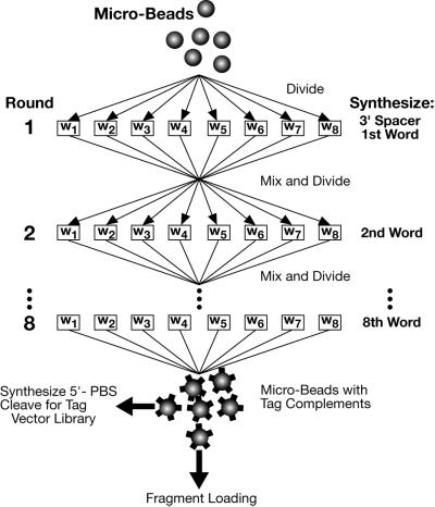
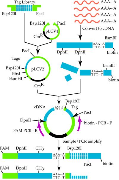
We describe a method for cloning nucleic acid molecules onto the surfaces of 5-micrometer microbeads rather than in biological hosts. A unique tag sequence is attached to each molecule, and the tagged library is amplified. Unique tagging of the molecules is achieved by sampling a small fraction (1%) of a very large repertoire of tag sequences. The resulting library is hybridized to microbeads that each carry approximately 10(6) strands complementary to one of the tags. About 10(5) copies of each molecule are collected on each microbead. Because such clones are segregated on microbeads, they can be operated on simultaneously and then assayed separately. To demonstrate the utility of this approach, we show how to label and extract microbeads bearing clones differentially expressed between two libraries by using a fluorescence-activated cell sorter (FACS). Because no prior information about the cloned molecules is required, this process is obviously useful where sequence databases are incomplete or nonexistent. More importantly, the process also permits the isolation of clones that are expressed only in given tissues or that are differentially expressed between normal and diseased states. Such clones then may be spotted on much more cost-effective, tissue- or disease-directed, low-density planar microarrays.


Figures

Figure 1
Figure 1
Tags were synthesized by eight rounds of combinatorial synthesis, wherein each word, w1–w8, is added in a separate column of a DNA synthesizer. In each round, a word was added to each growing tag by four conventional base-coupling cycles, after which the microbeads were mixed and divided for the next addition. After the eighth round, a portion of the microbeads were separated for further synthesis of a 5′ primer-binding site (PBS), followed by cleavage, amplification, and insertion into pLCV1.
Figure 2
Figure 2
Attachment of tags to cDNAs.
Figure 3
Figure 3
Melting curve of tags with complements. (A) Approximately 0.35 OD units of each double-stranded oligonucleotide was resuspended in 10 mM sodium phosphate, pH 7.6/50 mM NaCl/3 mM MgCl2. The samples were heated at 1°C per min, and the OD at 260 nm was measured and recorded. Reactions consisted of one common oligonucleotide, cccatcactttatcaatcaacatatcacaaaaatctcc, and a second oligonucleotide as follows: perfect match, (●) ggagatttttgtgatatgttgattgataaagtgatggg; one-word mismatch, (○) ggatgattttgtgatatgttgattgataaagtgatggg; two-word mismatch, (▵) ggagatttttgtttgatgttttgtgataaagtgatggg. (B) Microbeads bearing the oligonucleotide ggagatttttgtgatatgttgattgataaagtgatggg were loaded with FAM-labeled cDNA tagged with this sequence. In separate tubes, three cDNAs whose tags differed at the first, third, or fifth positions also were loaded. The samples then were washed in 50 mM Tris/50 mM NaCl/3 mM Mg2Cl at increasing temperatures as indicated. Microbeads then were analyzed by FACS, and the means of fluorescence intensities were plotted for the perfect match of the tag (filled bar), the mismatches (gray bars), and the ratios (cross-hatched bar) of the perfect signal to the average noise of the three mismatches.
Figure 4
Figure 4
Two-parameter, 530-nm (PMT1) and 666-nm (PMT2), FACS contour plot showing separation of loaded microbeads (contour on right) from unloaded microbeads (contour on left).
Figure 5
Figure 5
Model system for two-color competitive hybridization. A 34-mer, ggagatttgataaagtttgatgtgtaatagaggg, was synthesized with a 3′ FAM or Cy5 label. The oligonucleotides were brought to final concentration of 14 μM in 30 μl. The two labeled oligonucleotides were combined in nine separate mixtures in ratios of 1:0, 8:1, 4:1, 2:1, 1:1, 1:2, 1:4, 1:8, and 0:1. Hybridization of each mixture to 100,000 microbeads bearing the complementary oligonucleotide was performed, and 10,000 microbeads from each of the mixtures were analyzed with a Coulter Elite Flow Cytometer using 488-nm and 633-nm lasers.
Figure 6
Figure 6
A reference library of 2,000,000 microbeads was formed by mixing equal numbers of microbeads with attached cDNA derived from induced and noninduced THP-1 cells. Reference microbeads (100,000) were hybridized with 10 μg each of Cy5-labeled probe and R110-labeled probe, both derived from the same induced library to yield A. Microbeads (1,600,000) were hybridized with 10 μg of Cy5-labeled probe from the induced library and 10 μg of R110-labeled probe derived from the noninduced library to yield B.

References

    1. Antequera F, Bird A. Proc Natl Acad Sci USA. 1993;90:11995–11999. - PMC - PubMed
    1. Rowen L, Mahairas G, Hood L. Science. 1997;278:605–607. - PubMed
    1. Bishop J O, Morton J G, Rosbash M, Richardson M. Nature (London) 1974;250:199–204. - PubMed
    1. Ostrow R S, Woods W G, Vosika G J, Faras A J. Biochim Biophys Acta. 1979;562:92–102. - PubMed
    1. Lee C, Klopp R G, Weindruch R, Prolla T A. Science. 1999;285:1390–1393. - PubMed

MeSH terms

Associated data