. 2002 Apr 2;99(7):4471-6.
doi: 10.1073/pnas.072087599. Epub 2002 Mar 26.

Mutation of melanosome protein RAB38 in chocolate mice

Affiliations

Mutation of melanosome protein RAB38 in chocolate mice

Stacie K Loftus et al. Proc Natl Acad Sci U S A. .

Abstract

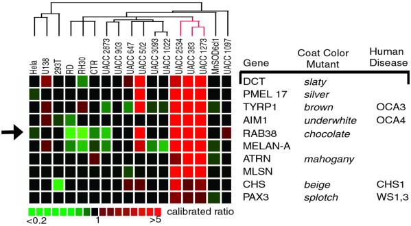
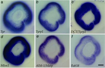
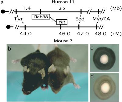
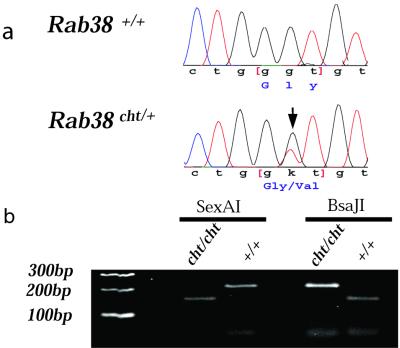
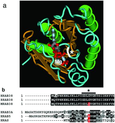
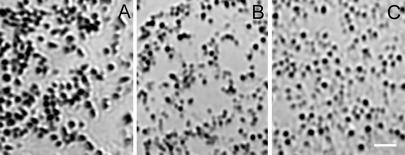
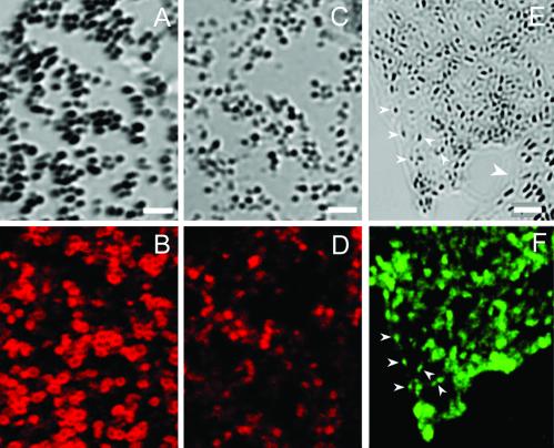
Mutations of genes needed for melanocyte function can result in oculocutaneous albinism. Examination of similarities in human gene expression patterns by using microarray analysis reveals that RAB38, a small GTP binding protein, demonstrates a similar expression profile to melanocytic genes. Comparative genomic analysis localizes human RAB38 to the mouse chocolate (cht) locus. A G146T mutation occurs in the conserved GTP binding domain of RAB38 in cht mice. Rab38(cht)/Rab38(cht) mice exhibit a brown coat similar in color to mice with a mutation in tyrosinase-related protein 1 (Tyrp1), a mouse model for oculocutaneous albinism. The targeting of TYRP1 protein to the melanosome is impaired in Rab38(cht)/Rab38(cht) melanocytes. These observations, and the fact that green fluorescent protein-tagged RAB38 colocalizes with end-stage melanosomes in wild-type melanocytes, suggest that RAB38 plays a role in the sorting of TYRP1. This study demonstrates the utility of expression profile analysis to identify mammalian disease genes.


Figures

Figure 1
Figure 1
Microarray expression profile of melanocyte control genes and RAB38. Expression profiles of nine genes known to be involved in pigmentation are similar to the expression profile of RAB38 (arrow). Columns to the right list identified mouse and human pigmentation disorders corresponding to a given gene. Hybridized sample cell lines are listed across the top of the expression profiles. Relative gene expression is evaluated as a calibrated ratio (sample cell line/MnSOD6cl1 reference). Pseudocolor scale for ratio values is below.
Figure 2
Figure 2
Rab38 expression in the RPE is similar to that of melanogenic enzymes. All panels show the eye at embryonic day 11.5. The melanogenic enzymes tyrosinase (Tyr) (a), tyrosinase-related protein 1 (Tyrp1) (b), and Dopachrome tautamerase (DCT/Tyrp2) (c) all show expression in the RPE at this developmental stage. Additional control genes from the microarray cluster data, Melastatin1 (Mlsn) (d) and AimI/Matp (e), show expression in the RPE. Rab38 is expressed in the RPE at embryonic day 11.5 (f) and at embryonic days 10.5 and 12.5 (data not shown). (Scale bar equals 100 μm.)
Figure 3
Figure 3
Map location and phenotype of cht/cht mouse. (a) Human RAB38 maps 1.4 Mb distal to tyrosinase and 2.5 Mb proximal to EED genes on human chromosome 11. In the corresponding mouse chromosome 7 syntenic region, gene order is conserved. The genetic location of the cht locus maps to this same interval as RAB38. (b) Photograph of C57BL/6J+/+ (Left, black) and C57BL/6J Rab38cht/Rab38cht (Right, brown) mice. Eyes from 2-day-old mice (c) wild-type C57Bl6/J+/+ shows normal pigmentation, whereas (d) Rab38cht/Rab38cht eye shows reduced pigmentation. (Magnification: ×50.)
Figure 4
Figure 4
Rab38 mutation causes the cht mouse phenotype. (a) Comparison of Rab38 sequence between wild-type C57BL/6J+/+ and C57BL/6J Rab38cht/+ DNA identified a G146T nucleotide change (arrow) for the cht allele. This nucleotide change was never seen in eight additional inbred strains analyzed (data not shown). (b) The G146T mutation creates a SexA1 restriction enzyme site in C57BL/6J Rab38cht/Rab38cht DNA and ablates a BsaJI restriction site present in wild-type Rab38 sequence. A 216-bp region surrounding the G146T nucleotide mutation was amplified from both C57BL/6J+/+ and C57BL/6J Rab38cht/Rab38cht DNA. SexA1 digests the PCR fragment of C57BL/6J Rab38cht/Rab38cht but not C57BL/6J+/+ (Left); BsaJ1 digests the PCR fragment of C57BL/6J+/+ but not C57BL/6J Rab38cht/Rab38cht (Right).
Figure 5
Figure 5
RAB38 G19 is located in the GTP binding pocket. (a) The three-dimensional location of amino acid G19 of RAB38 in relation to the nucleotide binding site was determined by using the molecular modeling database (MMDB) (40) based on the crystal structure for RAB3a (MMDB 10125) (34). Overlaying RAB38 sequence with that of RAB3a sequence identified amino acid S32 of RAB3a as equivalent to G19 of RAB38. The program CN3D 3.0 was used to indicate the location of the RAB38 G19 (white), predicting interaction with the bound nucleotide. Protein structure is indicated by color: green, α helices; gold, β sheet; blue, random coils; white, site of RAB3a S32 equivalent to RAB38 G19 located at the nucleotide binding site; gray, Mg++ ion; red-gray, GppNHp nucleotide analog. (b) Alignments of highly conserved N-terminal region comparing human RAB38 (NP_071732), rat RAB38 (AAA42000), and mouse RAB38 (AK009296.1). Corresponding alignments to human RAB3a (P20336), human RAB5 (F34323) and human N-RAS (TVHURA). Sequence alignment was done by using the clustalw algorithm (41). Bars indicate highly conserved regions that occupy the nucleotide binding pocket, demonstrated in the x-ray crystal structure of RAB3a (42). Black denotes sequence identity, gray denotes sequence conservation, red denotes the conserved amino acid.
Figure 6
Figure 6
The cht/cht melanosomes are similar in morphology to Tyrp1b melanosomes. Shown are bright-field images of melanosomes from the periphery of primary cultured melanocytes, isolated from (A) C57BL/6J+/+, (B) C57BL/6J Rab38cht/Rab38cht mice, and (C) Tyrp1b/Tyrp1b melan-b cells. Melanosomes from wild-type melanocytes are oval and darkly pigmented. In contrast, Rab38cht/Rab38cht melanosomes are smaller, more circular and less pigmented, similar to melanosomes from (C) Tyrp1b/Tyrp1b melan-b cells. (Scale bar = 2 μM.)
Figure 7
Figure 7
RAB38 is a melanosomal protein needed for appropriate TYRP1 trafficking. Shown are (A and B) C57BL/6J+/+ and (C and D) C57BL/6J Rab38cht/Rab38cht primary melanocyte cultures. (A and C) Bright-field images of melanosomes in the cell periphery with matching confocal images of identical exposure demonstrate distribution of (B and D) TYRP1 immunofluoresence as revealed by staining of MEL5. Melan-a cells transfected with a Rab38-GFP expression construct. (E) Bright-field image of melanosome cell periphery and (F) matching GFP fluorescence image for transfected cell. (E) Large white arrow indicates a nontransfected cell. (E and F) Small arrows demonstrate colocalization of Rab38-GFP signal with highly pigmented end stage melanosomes at cell periphery. (Scale bar: AD = 1.6 μM; E and F = 2.4 μM.)

References

    1. Jackson I J. Hum Mol Genet. 1997;6:1613–1624. - PubMed
    1. King R A, Hearing V J, Creel D, Oetting W S. In: The Metabolic Basis of Inherited Disease. 7th Ed. Scriver C R, Beaudet A L, Sly W S, Valle D V, editors. New York: McGraw–Hill; 1995. pp. 4353–4392.
    1. Newton J M, Cohen-Barak O, Hagiwara N, Gardner J M, Davisson M T, King R A, Brilliant M H. Am J Hum Genet. 2001;69:981–988. - PMC - PubMed
    1. Marks M S, Seabra M C. Nat Rev Mol Cell Biol. 2001;2:738–748. - PubMed
    1. Loftus S K, Pavan W J. Pigment Cell Res. 2000;13:141–146. - PubMed

Associated data