. 2003 Apr;71(4):1706-18.
doi: 10.1128/IAI.71.4.1706-1718.2003.

Expression of multiple outer membrane protein sequence variants from a single genomic locus of Anaplasma phagocytophilum

Affiliations

Expression of multiple outer membrane protein sequence variants from a single genomic locus of Anaplasma phagocytophilum

A F Barbet et al. Infect Immun. 2003 Apr.

Abstract

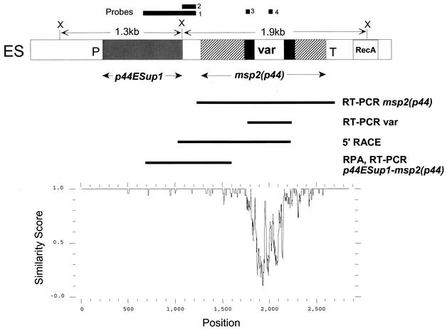
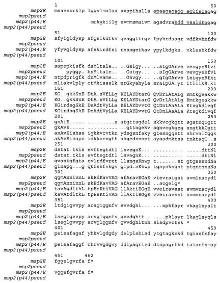
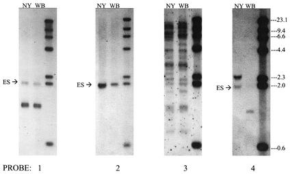
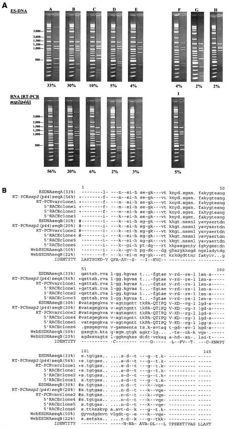
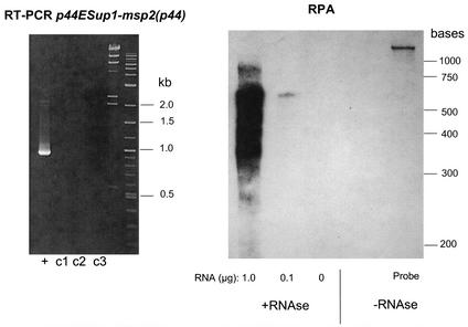
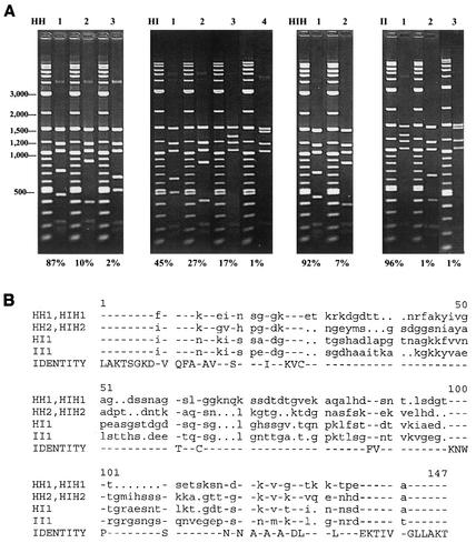
Anaplasma phagocytophilum is the causative agent of an emerging tick-borne zoonosis in the United States and Europe. The organism causes a febrile illness accompanied by other nonspecific symptoms and can be fatal, especially if treatment is delayed. Persistence of A. phagocytophilum within mammalian reservoir hosts is important for ensuring continued disease transmission. In the related organism Anaplasma marginale, persistence is associated with antigenic variation of the immunoprotective outer membrane protein MSP2. Extensive diversity of MSP2 is achieved by combinatorial gene conversion of a genomic expression site by truncated pseudogenes. The major outer membrane protein of A. phagocytophilum, MSP2(P44), is homologous to MSP2 of A. marginale, has a similar organization of conserved and variable regions, and is also encoded by a multigene family containing some truncated gene copies. This suggests that the two organisms could use similar mechanisms to generate diversity in outer membrane proteins from their small genomes. We define here a genomic expression site for MSP2(P44) in A. phagocytophilum. As in A. marginale, the msp2(p44) gene in this expression site is polymorphic in all populations of organisms we have examined, whether organisms are obtained from in vitro culture in human HL-60 cells, from culture in the tick cell line ISE6, or from infected human blood. Changes in culture conditions were found to favor the growth and predominance of certain msp2(p44) variants. Insertions, deletions, and substitutions in the region of the genomic expression site encoding the central hypervariable region matched sequence polymorphisms in msp2(p44) mRNA. These data suggest that, similarly to A. marginale, A. phagocytophilum uses combinatorial mechanisms to generate a large array of outer membrane protein variants. Such gene polymorphism has profound implications for the design of vaccines, diagnostic tests, and therapy.


Figures

FIG. 1.
FIG. 1.
Structure and variability of a genomic expression site for msp2(p44) in A. phagocytophilum. (Top) Diagram indicating the location of msp2(p44) and an upstream gene (p44ESup1) within the expression site. Solid areas immediately flanking the variable region (var) represent sequence present in the expression site and in most genomic pseudogenes. The locations of RT-PCR and 5′-RACE products and of an RPA probe used to establish the structure of mRNA carrying msp2(p44) are indicated below the diagram. DNA probes used in Southern blots to verify locus structure are indicated above the diagram. ES, expression site; P, promoter sequence; T, terminator; RecA, a downstream sequence homologous to the recA gene; X, XbaI cleavage site. (Bottom) PLOTSIMILARITY graph, drawn to the same scale as the diagram above, demonstrating the variability of this expression locus in five different populations of A. phagocytophilum. These populations are the NY18 strain cultured in HL-60 cells, the Webster strain cultured in HL-60 cells, the HGE2 strain cultured in ISE6 tick cells (population II in Fig. 7), and two populations of A. phagocytophilum from infected human blood (patient 2, day 3, and patient 2, day 27 [Fig. 8]). A similarity score of 1.0 indicates identical sequence in a sliding window of 10 nucleotides, and a score decreasing from 1.0 to 0.0 indicates increasing variation.
FIG. 2.
FIG. 2.
Conservation of msp2 sequence between A. marginale and A. phagocytophilum. The msp2E sequence is the predominant variant sequence encoded in the msp2 expression site of an acute bloodstream population of A. marginale strain Florida (GenBank accession number AF200925), and msp2pseud is encoded by a pseudogene present in genomic DNA of the same strain of A. marginale (accession number U60780). The msp2(p44)E sequence is the predominant variant sequence encoded in the msp2(p44) expression site of A. phagocytophilum strain NY18 (this study) grown in HL-60 cells, and msp2(p44)pseud is encoded by a pseudogene present in genomic DNA of the same strain of A. phagocytophilum (this study). Amino acids that are identical in all four sequences are capitalized. The N-terminal amino acid sequences of native MSP2 and MSP2(P44) are underlined.
FIG. 3.
FIG. 3.
Verification of expression site structure by Southern blotting of genomic DNA. A. phagocytophilum DNA from either strain NY18 (NY) or strain Webster (WB) was digested with the enzyme XbaI to release the 1.9-kb fragment containing the expressed msp2(p44) gene (ES) and the 1.3-kb fragment containing p44ESup1. Digested and separated DNA was hybridized with probes 1 to 4 against different regions of the expression site (see Fig. 1 for locations of probes). Molecular weight markers are in the far right lane of each blot.
FIG. 4.
FIG. 4.
Comparison of outer membrane protein expression site structures in A. marginale and A. phagocytophilum. (A) Diagram showing the locations of expressed msp2 and msp2(p44) genes in the two organisms. Nomenclature for opag1 to opag3 of A. marginale is as in reference . P, predicted promoter; T, predicted prokaryotic terminator sequence; var, variable region. (B) Comparison of predicted promoter sequences in A. marginale and A. phagocytophilum expression sites.
FIG.5.
FIG.5.
Sequence diversity in mRNA encoding msp2(p44) reflects polymorphisms in the msp2(p44) copy within the genomic expression site. (A) RFLP analysis of expression site clones compared to RFLP analysis of msp2(p44) mRNA. Plasmid DNAs from 135 independent strain NY18 genomic expression site clones (ES-DNA) and 95 independent clones derived by RT-PCR from NY18 msp2(p44) mRNA were digested with EcoRI and RsaI and analyzed by agarose gel electrophoresis. Clones with identical digestion patterns were grouped together, and the frequencies of the major patterns were determined. Each digestion pattern (on the right) is shown next to a marker lane of molecular weight standards. The percentage of clones with each digestion pattern is shown below. Minor patterns representing <2% of the population are not shown; therefore, the sums of the percentages shown are <100%. The predominant variants, represented by clones A through E, have the same digestion patterns whether they are derived from DNA or from RNA. Patterns F through I, representing minor variants, were unique to clones derived from either DNA or RNA. (B) Sequence comparison of genomic expression site clones with clones derived from msp2(p44) mRNA. Individual expression site clones representing the predominant RFLP patterns A and B (Fig. 5A) were sequenced and aligned with the sequences of six 5′-RACE clones and two RT-PCRvar clones, also derived from strain NY18 msp2(p44) mRNA. For comparison, the two predominant genomic expression site sequence variants present in strain Webster (WebESDNAseqA and WebESDNAseqB) are included at the bottom of the alignment. The amino acids encoded by probe 4 (see Fig. 1 and 3), specific for the CVR sequence expressed in strain NY18, are capitalized (amino acids 73 to 84). Identical sequences are indicated by identical symbols (+ or #) to the left of the aligned sequences. Although not identical, the sequences of ESDNAseqA and 5′-RACE clone 5 each differed by only a single amino acid from the two groups of identical sequence variants. These changes could represent actual minor variation in mRNA species or a mutation occurring during in vitro amplification and cloning.
FIG. 6.
FIG. 6.
The major mRNA species encoding the N terminus of msp2(p44) in A. phagocytophilum-infected HL-60 cells also contains the msp2(p44)-p44ESup1 intergenic region. (Left) Ethidium bromide-stained gel of RT-PCRs amplifying the C-terminal region encoding p44ESup1, the N-terminal region encoding msp2(p44), and the intergenic region between them. +, RT-PCR; c1 to c3, control reactions containing either no reverse transcriptase enzyme, no template in primary PCR, or no template in secondary PCR, respectively. (Right) RPA using the cloned and sequenced 907-bp RT-PCR product to generate an antisense RNA probe. The complete RNA probe is ∼1,100 bases, as it also contains vector sequence that should not be protected by hybridization with A. phagocytophilum RNA. The quantity of protecting A. phagocytophilum RNA is indicated below the gel. Positions of molecular size standards are indicated on the right for both panels.
FIG. 7.
FIG. 7.
The predominant msp2(p44) sequence variants in the expression site are different during in vitro growth in HL-60 and ISE6 cells. A. phagocytophilum was grown continuously in HL-60 (HH) or ISE6 (II) cells or transferred between them (HI, HIH). A total of 70 to 92 independent clones of the genomic expression site were prepared and analyzed from each of the four populations of A. phagocytophilum, as in Fig. 5. (A) RFLP analysis of expression site clones from each population. The percentage of clones with each digestion pattern is given below each gel. (B) Alignment of the CVRs of the msp2(p44) expression sites from the predominant sequence variants determined in panel A.
FIG. 8.
FIG. 8.
A diverse repertoire of msp2(p44) sequences is encoded in the genomic expression site in individual patients infected with A. phagocytophilum. Genomic DNA was prepared from infected human blood of each patient. For two patients (patients 1 and 2), samples were available at differing times following the first onset of clinical symptoms. Independent clones of the genomic expression site were prepared and analyzed by RFLP mapping as in Fig. 5 and 7. The predominant variants (A and B) in each sample were sequenced and aligned by using PILEUP. In one case a minor variant in one population (patient 2, day 3, varC) that was identical to a predominant variant in a subsequent A. phagocytophilum population from the same patient (patient 2, day 27, varA) is also shown. The percentage of each sequence variant within a population, as determined by RFLP mapping, is indicated next to the variant designation. Identical sequences are indicated by identical symbols (+, #, or x) to the left of aligned sequences.

References

    1. Aguero, R. M., H. W. Horowitz, G. P. Wormser, D. F. McKenna, J. Nowakowski, J. Munoz, and J. S. Dumler. 1996. Human granulocytic ehrlichiosis: a case series from a medical center in New York State. Ann. Intern. Med. 125:904-908. - PubMed
    1. Asanovich, K. M., J. S. Bakken, J. E. Madigan, R. M. Aguero, G. P. Wormser, and J. S. Dumler. 1997. Antigenic diversity of granulocytic Ehrlichia isolates from humans in Wisconsin and New York and a horse in California. J. Infect. Dis. 176:1029-1034. - PubMed
    1. Barbet, A. F., A. Lundgren, J. Yi, F. R. Rurangirwa, and G. H. Palmer. 2000. Antigenic variation of Anaplasma marginale by expression of MSP2 mosaics. Infect. Immun. 68:6133-6138. - PMC - PubMed
    1. Brayton, K. A., D. P. Knowles, T. C. McGuire, and G. H. Palmer. 2001. Efficient use of a small genome to generate diversity in tick-borne ehrlichial pathogens. Proc. Natl. Acad. Sci. USA 98:4130-4135. - PMC - PubMed
    1. Brayton, K. A., G. H. Palmer, A. Lundgren, J. Yi, and A. F. Barbet. 2002. Antigenic variation of Anaplasma marginale msp2 occurs by combinatorial gene conversion. Mol. Microbiol. 43:1151-1159. - PubMed

Publication types

MeSH terms

Substances

Associated data