. 2003 Feb;13(2):182-94.
doi: 10.1101/gr.681703.

Sequence analysis of a functional Drosophila centromere

Affiliations

Sequence analysis of a functional Drosophila centromere

Xiaoping Sun et al. Genome Res. 2003 Feb.

Abstract

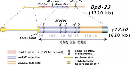
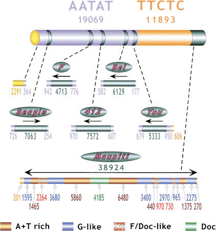
Centromeres are the site for kinetochore formation and spindle attachment and are embedded in heterochromatin in most eukaryotes. The repeat-rich nature of heterochromatin has hindered obtaining a detailed understanding of the composition and organization of heterochromatic and centromeric DNA sequences. Here, we report the results of extensive sequence analysis of a fully functional centromere present in the Drosophila Dp1187 minichromosome. Approximately 8.4% (31 kb) of the highly repeated satellite DNA (AATAT and TTCTC) was sequenced, representing the largest data set of Drosophila satellite DNA sequence to date. Sequence analysis revealed that the orientation of the arrays is uniform and that individual repeats within the arrays mostly differ by rare, single-base polymorphisms. The entire complex DNA component of this centromere (69.7 kb) was sequenced and assembled. The 39-kb "complex island" Maupiti contains long stretches of a complex A+T rich repeat interspersed with transposon fragments, and most of these elements are organized as direct repeats. Surprisingly, five single, intact transposons are directly inserted at different locations in the AATAT satellite arrays. We find no evidence for centromere-specific sequences within this centromere, providing further evidence for sequence-independent, epigenetic determination of centromere identity and function in higher eukaryotes. Our results also demonstrate that the sequence composition and organization of large regions of centric heterochromatin can be determined, despite the presence of repeated DNA.


Figures

Figure 1.
Figure 1.
Origin and structure of γ1230. γ1230 was generated by radiation-induced deletions of Dp823(Le et al. 1995). The positions of the “islands” of complex DNA (Tahiti, Moorea, Bora Bora, and Maupiti) are shown, as is the position of the new euchromatin/heterochromatin junction (“breakpoint”). The 420-kb functional centromere was localized by examining the transmission behavior of a large collection of minichromosome derivatives (Murphy and Karpen 1995b). Restriction mapping and hybridization analysis indicated that the centromere portion of γ1230 contained two satellite DNA blocks (AATAT and AAGAG), five small islands of complex DNA (Motus) inserted into the AATAT block, and a large region of complex DNA (Maupiti) at the right end (Sun et al. 1997). The sizes of the satellite blocks are shown.
Figure 2.
Figure 2.
Summary of γ1230 centromeric sequences. Numbers below AATAT and TTCTC report the total amount of sequence (base pairs) generated for each satellite, including blocks flanking complex DNA as well as the random, unmapped sequences generated by tagged PCR. Numbers below each transposon “bar” and the Maupiti diagram report the amount of contiguous sequence generated for each region and type of DNA, in base pairs. Arrows indicate the 5′ to 3′ orientation of the transposons, relative to previously sequenced euchromatic elements.
Figure 3.
Figure 3.
“Tagged” polymerase chain reaction (PCR) methods for cloning and sequencing heterochromatin. (A) Standard PCR amplifications with one satellite primer and one “unique” primer (homologous to the end of the transposon) result in shrinkage with each round of amplification. A satellite repeat primer will anneal anywhere in the repeated template, not just at the ends, and thus successive rounds of amplification will result in shorter and shorter products. The template shrinkage problem is even greater when two satellite primers are used to amplify pure satellite arrays (not shown), and in addition, the self-complementarity of the two satellite primers results in primer-dimer formation (not shown). (B) Junctions between complex DNA (transposons in this case) and satellite arrays are successfully amplified with significantly less shrinkage when a hybrid GC-tagged/satellite primer is used. Initial amplification at low stringency incorporates the tag at random within the satellite array then extends across the location of the TE primer; the subsequent exponential amplification reduces shrinkage because high stringency mandates annealing of the entire hybrid primer. This method was also used to generate sequence from bacterial transposon insertions into gel-purified γ1230; in this case, the “complex” primer was homologous to the end of the bacterial transposon. (C) Pure satellite arrays were amplified with two tagged primers, plus a primer corresponding to one of the tags. One tagged primer was used for single-stranded extension at low stringency; after gel isolation of the single-strand products, the second primer was used to synthesize the complementary strand, then the tag and one tagged primer were used for high stringency amplification.
Figure 3.
Figure 3.
“Tagged” polymerase chain reaction (PCR) methods for cloning and sequencing heterochromatin. (A) Standard PCR amplifications with one satellite primer and one “unique” primer (homologous to the end of the transposon) result in shrinkage with each round of amplification. A satellite repeat primer will anneal anywhere in the repeated template, not just at the ends, and thus successive rounds of amplification will result in shorter and shorter products. The template shrinkage problem is even greater when two satellite primers are used to amplify pure satellite arrays (not shown), and in addition, the self-complementarity of the two satellite primers results in primer-dimer formation (not shown). (B) Junctions between complex DNA (transposons in this case) and satellite arrays are successfully amplified with significantly less shrinkage when a hybrid GC-tagged/satellite primer is used. Initial amplification at low stringency incorporates the tag at random within the satellite array then extends across the location of the TE primer; the subsequent exponential amplification reduces shrinkage because high stringency mandates annealing of the entire hybrid primer. This method was also used to generate sequence from bacterial transposon insertions into gel-purified γ1230; in this case, the “complex” primer was homologous to the end of the bacterial transposon. (C) Pure satellite arrays were amplified with two tagged primers, plus a primer corresponding to one of the tags. One tagged primer was used for single-stranded extension at low stringency; after gel isolation of the single-strand products, the second primer was used to synthesize the complementary strand, then the tag and one tagged primer were used for high stringency amplification.
Figure 4.
Figure 4.
Sequence composition and organization of Maupiti. (A) Dot-plot analysis of Maupiti reveals the presence and organization of internally repeated DNAs. Arrows above each bar indicate relative orientations of each component. Solid arrows below the bars indicate the presence of inverted repeats at the ends, and large internal direct repeats. Note that most of the internal elements are oriented in the same direction. (B) The substructures of the A+T-rich, G-like, and Doc elements are shown relative to each other, and to previously sequenced elements (bottom). Blocks are numbered from left to right (relative to A). The homologies (colors) and relative orientations (arrowheads) of subrepeats within the A+T-rich elements are shown. The inverted repeats at the ends are composed of G-like block 1/A+T-rich block 1 and A+T-rich block 5/G-like block 6.

References

    1. The Arabidopsis Genome Initiative, 2000. Analysis of the genome sequence of the flowering plant Arabidopsis thaliana. In Nature, pp. 796–815. - PubMed
    1. Adams M.D., Celniker, S.E., Holt, R.A., Evans, C.A., Gocayne, J.D., Amanatides, P.G., Scherer, S.E., Li, P.W., Hoskins, R.A., Galle, R.F., et al. 2000. The genome sequence of Drosophila melanogaster. Science 287: 2185-2195. - PubMed
    1. Allshire R.C., Javerzat, J.P., Redhead, N.J., and Cranston, G. 1994. Position effect variegation at fission yeast centromeres. Cell 76: 157-169. - PubMed
    1. Arn P.H., Li, X., Smith, C., Hsu, M., Schwartz, D.C., and Jabs, E.W. 1991. Analysis of DNA restriction fragments greater than 5.7 Mb in size from the centromeric region of human chromosomes. Mamm. Genome 1: 249-254. - PubMed
    1. Barry A.E., Bateman, M., Howman, E.V., Cancilla, M.R., Tainton, K.M., Irvine, D.V., Saffery, R., and Choo, K.H. 2000. The 10q25 neocentromere and its inactive progenitor have identical primary nucleotide sequence: Further evidence for epigenetic modification. Genome Res. 10: 832-838. - PMC - PubMed

Publication types

MeSH terms

Associated data

LinkOut - more resources