. 2003 May 13;100(10):6081-6.
doi: 10.1073/pnas.0937739100. Epub 2003 Apr 25.

Adeno-associated viruses undergo substantial evolution in primates during natural infections

Affiliations

Adeno-associated viruses undergo substantial evolution in primates during natural infections

Guangping Gao et al. Proc Natl Acad Sci U S A. .

Abstract

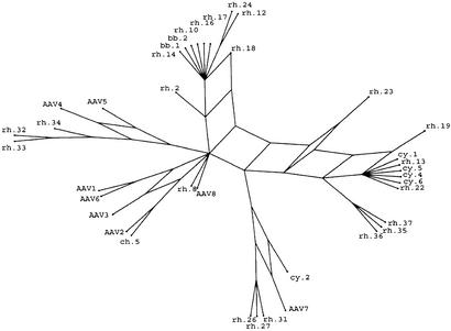
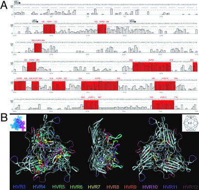
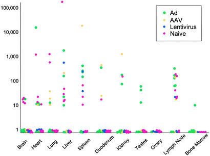
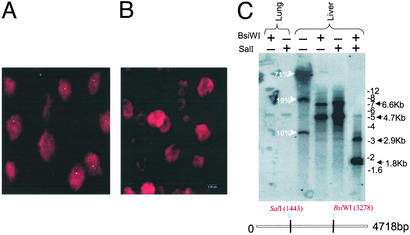
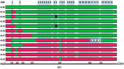
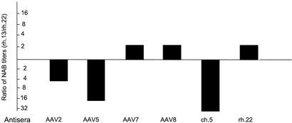
Adeno-associated viruses (AAVs) are single-stranded DNA viruses that are endemic in human populations without known clinical sequelae and are being evaluated as vectors for human gene therapy. To better understand the biology of this virus, we examined a number of nonhuman primate species for the presence of previously uncharacterized AAVs and characterized their structure and distribution. AAV genomes were widely disseminated throughout multiple tissues of a variety of nonhuman primate species. Surprising diversity of sequence, primarily localized to hypervariable regions of the capsid protein, was detected. This diversity of sequence is caused, in part, by homologous recombination of co-infecting parental viruses that modify the serologic reactivity and tropism of the virus. This is an example of rapid molecular evolution of a DNA virus in a way that was formerly thought to be restricted to RNA viruses.


Figures

Figure 1
Figure 1
Split-decomposition analysis of 38 AAV sequences. The split-decomposition method performed as described in Materials and Methods represents the previously uncharacterized AAVs in relation to the known serotypes. This model is not on scale and magnifies network links from the analysis for better presentation.
Figure 2
Figure 2
Location of HVRs on both the primary and tertiary capsid structure of AAV2. (A) A total of 12 HVRs were identified and are highlighted in red on the figure in combination with their respective AAV2 coordinates. Plotted along the x axis are the coordinates of the 38 VP1 protein sequence alignments in combination with the AAV2 residue. Gaps in the AAV2 sequence are denoted with X. The y axis represents 100 − Q, an indicator of variation in the alignment. More variation is represented by a higher number. The black arrows indicate the N terminus of the different capsid proteins. (B) Ten HVRs (HVRs 3–12) of the AAV capsid protein are located on the resolved x-ray structure of the AAV2 capsid. Each capsid facet contains three subunits, one of which is highlighted (Upper Left; PDB ID code 1LP3). (Upper Right) A schematic shows the relative orientation of the 20 capsid facets and the five-, three-, and twofold symmetry elements. A ribbon drawing of facet with HVRs colored as viewed from the outside (Left), edge (Center), and inside (Right) of the virus. All these HVRs are located on the surface of the virion, and most are clustered in the threefold-related towers.
Figure 3
Figure 3
Tissue distribution of AAV sequences in rhesus macaques. Genome copies of AAV sequences present in DNA samples extracted from 10 different tissues of 21 rhesus macaques that had not been enrolled in a previous study or had been administered an AAV, adenovirus (Ad), or lentiviral vector as part of another study were quantified by real-time PCR. Copy numbers of AAV genomes are reported as AAV copies per 100 ng of macaque genomic DNA detected in a 50-cycle amplification (y axis). The x axis represents different tissues surveyed in the study. The history of each animal is illustrated by the color of solid dots. The animals were naive at the time of necropsy (i.e., not on study, shown in pink) or were being necropsied after exposure to adenoviral (green), AAV (yellow), or lentiviral (blue) vectors.
Figure 4
Figure 4
Molecular characterization and cellular distribution of AAV sequences in tissues. (A and B) In situ hybridization was performed on liver sections from rhesus macaques by using a digoxigenin-labeled AAV8 probe, and optical sections were collected at 0.5-μm intervals. A digital merge of individual green (digoxigenin) and red (propidium iodide) channel images from midplane sections of animals with >20 (RQ4407, A) and <0.1 (V383, B) proviral copies per diploid genome is shown. (C) Molecular state of AAV sequences in the cellular DNA from liver and lung tissues of rhesus macaque RQ4407 was analyzed by DNA hybridization. Total cellular DNA was analyzed by DNA hybridization as described in Materials and Methods. The enzymes used are indicated, and the probe was a mixture of Rep and Cap sequences.
Figure 5
Figure 5
Structure of multiple AAV variants from a mesenteric lymph node of a rhesus macaque. The conserved regions were deleted from the alignment of the protein sequences of 12 AAV VP1 variants isolated from the mesenteric lymph node of animal Tulane/F953. The remaining variable residues were color-coded either red or green corresponding to their putative parental clone, rh.13 or rh.20, respectively. Location of the HVRs in relation to these differences are shaded in gray and numbered along the top. Each block represents a single residue or a group of neighboring residues. Black residues indicate a mutation that could not be brought back to any of the putative parents. X represents a deletion. Because of the nature of the presentation the coordinates corresponding to AAV2 VP1 amino acid residues are not linear.
Figure 6
Figure 6
Functional analysis of previously uncharacterized AAVs as pseudotypes. Example of the impact of capsid variation on serologic crossreactivity. Two pseudotypes from the mesenteric lymph nodes, which presumably are derived from recombination, were screened for neutralization against nonspecific antisera generated to a number of AAVs. Sera that show differences between pseudotypes based on rh.13 and rh.22 are presented as the ratios of neutralizing titers. Similar studies with sera from AAV1, rh.8, rh.10, rh.13, rh.21, and rh.24 showed no differences in neutralizing activity against rh.13 and rh.22. NAB, neutralizing antibody.

References

    1. Muzyczka N, Berns K I. In: Fields Virology. Knipe D M, Howley P M, editors. Vol. 2. Williams & Wilkins, Philadelphia: Lippincott; 2001. pp. 2327–2359.
    1. Kotin R M. Hum Gene Ther. 1994;5:793–801. - PubMed
    1. Parks W P, Boucher D W, Melnick J L, Taber L H, Yow M D. Infect Immun. 1970;2:716–722. - PMC - PubMed
    1. Atchison R W, Casto B C, Hammon W M. Science. 1965;149:754–756. - PubMed
    1. Mayor H D, Melnick J L. Nature. 1966;210:331–332. - PubMed

Publication types

MeSH terms

Associated data

LinkOut - more resources