. 2003 Oct;185(20):6083-94.
doi: 10.1128/JB.185.20.6083-6094.2003.

Global mutational analysis of NtrC-like activators in Myxococcus xanthus: identifying activator mutants defective for motility and fruiting body development

Affiliations

Global mutational analysis of NtrC-like activators in Myxococcus xanthus: identifying activator mutants defective for motility and fruiting body development

Nora B Caberoy et al. J Bacteriol. 2003 Oct.

Abstract

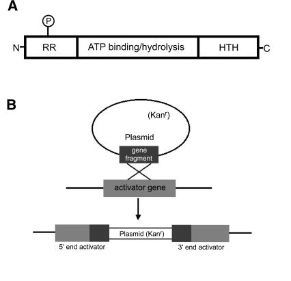
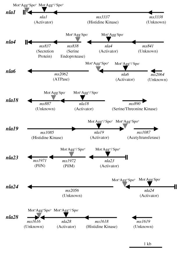
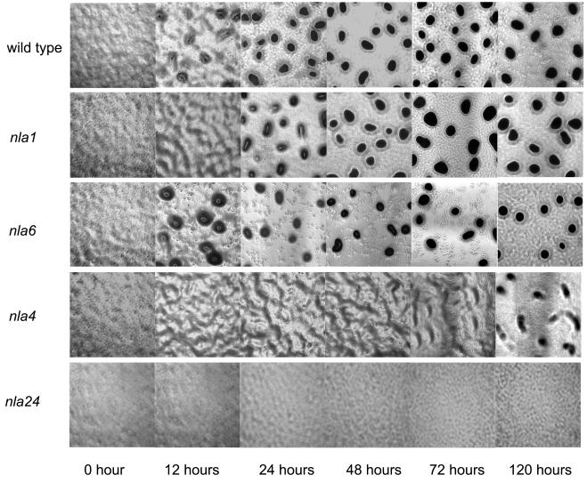
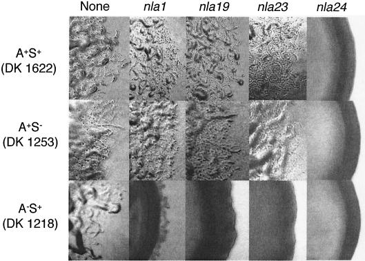
The multicellular developmental cycle of Myxococcus xanthus requires large-scale changes in gene transcription, and recent findings indicate that NtrC-like activators play a prominent role in regulating these changes. In this study, we made insertions in 28 uncharacterized ntrC-like activator (nla) genes and found that eight of these insertions cause developmental defects. Hence, these results are consistent with the idea that M. xanthus uses a series of different NtrC-like activators during fruiting body development. Four of the eight developmental mutants we identified have motility defects. The nla1, nla19, and nla23 mutants show S-motility defects, while the nla24 mutant shows defects in both S-motility and A-motility. During development, aggregation of the nla1, nla19, and nla23 mutants is delayed slightly and the nla24 mutant shows no signs of aggregation or sporulation. The nla4, nla6, nla18, and nla28 mutants have no appreciable loss in motility, but they fail to aggregate and to sporulate normally. The nla18 mutant belongs to a special class of developmental mutants whose defects can be rescued when they are codeveloped with wild-type cells, suggesting that nla18 fails to produce a cell-cell signal required for development. The three remaining activator mutants, nla4, nla6, and nla28, appear to have complex developmental phenotypes that include deficiencies in cell-cell developmental signals.


Figures

FIG. 1.
FIG. 1.
(A) Common structure of NtrC-like activators. The N-terminal domain is the least conserved of the domains indicated, and its length ranges from about 12 to 400 amino acids. Many activators serve as response regulators in two-component regulatory systems, as indicated by the letters RR. In the two-component paradigm, a histidine kinase sensor modulates the activity of a response regulator partner by phosphorylation of a conserved aspartate residue (P). The C-terminal domain of activators is about 65 to 130 amino acids. This region contains a helix-turn-helix (HTH) motif, which is characteristic of many DNA binding proteins. The central domain of activators is the most highly conserved region, and it consists of approximately 240 amino acids. This central domain is required for ATP binding and hydrolysis, which helps σ54-bound RNA polymerase become transcriptionally active. The central region of the activators may also be involved in contacting σ54-RNA polymerase. Data for this figure are taken from work by Morett and Segovia (46) and Xu and Hoover (66). (B) Disruption of ntrC-like activator (nla) genes by homologous recombination. Internal fragments of activator genes were cloned into a plasmid vector that confers resistance to kanamycin. After electroporation of the plasmid clones into wild-type M. xanthus cells, a single homologous crossover produces a tandem duplication of the internal fragment and incorporation of the vector into the chromosomal copy of the gene. The likely result of the crossover is an inactivated (knockout) copy of the activator gene.
FIG.2.
FIG.2.
Physical map of nla loci implicated in development. Gene designations were taken from the work of R. D. Welch, J. S. Jakobsen, and D. Kaiser (personal communication). The potential functions of some of the gene products are indicated in parentheses below the gene designations. “Unknown” indicates that in BLAST searches, no potential function for a gene product was obtained. Black inverted triangles represent insertions within nla genes, and gray inverted triangles represent insertions downstream of nla genes. The motility (Mot), aggregation (Agg), and sporulation (Spo) phenotypes produced by each insertion are indicated above the insertions. The data were taken from the results shown in Table 1, Table 2, and Table 4. Double bars (||) at the end of a line indicate that the upstream or downstream sequence was not available.
FIG. 3.
FIG. 3.
Behavior of representative nla mutants during development on TPM agar plates. Cells were spotted on TPM starvation agar and monitored visually as described in Materials and Methods. Development of the indicated nla mutants and wild-type strain DK1622 was observed for 5 days using a phase-contrast microscope. The aggregation phenotypes of nla19 and nla23 mutant cells are similar to those of nla1 mutant cells, and the aggregation phenotypes of nla18 and nla28 mutant cells are similar those of nla6 mutant cells. Photographs were taken after 12, 24, 48, 72, and 120 h using a total magnification of ×40.
FIG. 4.
FIG. 4.
Colony edge morphologies produced by nla insertions. The nla1, nla19, nla23, and nla24 insertions were transferred into A+ S+ (DK1622), A S+ (DK1218), and A+ S (DK1253) backgrounds, and colony edges were observed using phase-contrast microscopy (×40 magnification). Photographs were taken after 5 days on CTTYE plates.

References

    1. Arnold, J. W., and L. J. Shimkets. 1988. Inhibition of cell-cell interactions in Myxococcus xanthus by Congo red. J. Bacteriol. 170:5765-5770. - PMC - PubMed
    1. Arnold, J. W., and L. J. Shimkets. 1988. Cell surface properties correlated with cohesion in Myxococcus xanthus. J. Bacteriol. 170:5771-5777. - PMC - PubMed
    1. Behmlander, R. M., and M. Dworkin. 1994. Biochemical and structural analyses of the extracellular matrix fibrils of Myxococcus xanthus. J. Bacteriol. 176:6295-6303. - PMC - PubMed
    1. Brandner, J. P., and L. Kroos. 1998. Identification of the Ω4400 regulatory region, a developmental promoter of Myxococcus xanthus. J. Bacteriol. 180:1995-2004. - PMC - PubMed
    1. Bretscher, A. P., and D. Kaiser. 1978. Nutrition of Myxococcus xanthus, a fruiting myxobacterium. J. Bacteriol. 133:763-768. - PMC - PubMed

Publication types

Associated data

LinkOut - more resources