. 2005 Mar 15;102(11):4146-51.
doi: 10.1073/pnas.0407918102. Epub 2005 Mar 7.

A spatially localized rhomboid protease cleaves cell surface adhesins essential for invasion by Toxoplasma

Affiliations

A spatially localized rhomboid protease cleaves cell surface adhesins essential for invasion by Toxoplasma

Fabien Brossier et al. Proc Natl Acad Sci U S A. .

Abstract

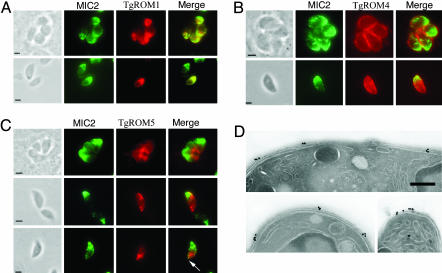
Apicomplexan parasites cause serious human and animal diseases, the treatment of which requires identification of new therapeutic targets. Host-cell invasion culminates in the essential cleavage of parasite adhesins, and although the cleavage site for several adhesins maps within their transmembrane domains, the protease responsible for this processing has not been discovered. We have identified, cloned, and characterized the five nonmitochondrial rhomboid intramembrane proteases encoded in the recently completed genome of Toxoplasma gondii. Four T. gondii rhomboids (TgROMs) were active proteases with similar substrate specificity. TgROM1, TgROM4, and TgROM5 were expressed in the tachyzoite stage responsible for the disease, whereas TgROM2 and TgROM3 were expressed in the oocyst stage involved in transmission. Although both TgROM5 and TgROM4 localized to the cell surface in tachyzoites, TgROM5 was primarily at the posterior of the parasite, whereas adhesins were sequestered in internal micronemes. Upon microneme secretion, as occurs during invasion, the MIC2 adhesin was secreted to the apical end and translocated to the posterior, the site of cleavage, where it colocalized only with TgROM5. Moreover, only TgROM5 was able to cleave MIC adhesins in a cell-based assay, indicating that it likely provides the key protease activity necessary for invasion. T. gondii rhomboids have clear homologues in other apicomplexans including malaria; thus, our findings provide a model for studying invasion by this deadly pathogen and offer a target for therapeutic intervention.


Figures

Fig. 1.
Fig. 1.
T. gondii contains five nonmitochondrial rhomboids, all of which are closely related to the Rho members of the family. (A) Schematic representation of the predicted T. gondii rhomboids. The seven TMDs (gray bars) are numbered. The three conserved catalytic triad residues (N in TMD2, the catalytic S in TMD4, and H in TMD6) and the GAST or GSSG motifs surrounding the putative active serines are indicated. (B) Unrooted phylogram based on neighbor-joining analysis of selected rhomboid proteases. Toxoplasma rhomboids grouped with orthologues in Plasmodium spp. were not closely aligned with other major groups of rhomboids found in bacteria, plants, or animals, thus forming several separate parasite groups. Bootstrap values are shown with colored circles on the tree corresponding the values given in the lower right corner. Dm, D. melanogaster; Hs, Homo sapiens; Mm, Mus musculus; Tg, T. gondii; A. thaliana, Arabidopsis thaliana; O. sativa, Oryza sativa; P. syringae, Pseudomonas syringae; AarA, Providencia stuartii; R. sphaerodies, Rhodobacter sphaerodies (GenBank accession numbers are beneath each entry).
Fig. 2.
Fig. 2.
Activity analysis of TgROMs. (A) Plasmids encoding GFP-Spitz and Star, the transport factor for Spitz (26), were cotransfected into COS cells alone (–), with 100 ng or 250 ng of a plasmid encoding one of the TgROMs, or 100 ng of Drosophila Rho-1 (Dm) as a positive control. When Spitz is cleaved, it is detected by anti-GFP Western blot analysis as a smaller band in cells (arrowhead in A and B) and as a secreted product in cell culture media. All TgROMs were tagged at their extreme N termini with a triple HA tag to verify their expression levels in mammalian cells [tagged forms retained wild-type (WT) levels of activity compared with untagged forms]. Standards in kDa are shown to the right of each panel. High amounts of TgROM5 (250 ng) resulted in cytotoxicity, which was also apparent as a reduction of Spitz in cells. Note that when cleavage occurs in the endoplasmic reticulum [Dm Rho-1-KDEL (R1K)], the cleaved product accumulates in cells and is poorly secreted (21) This observation might explain why the cleaved product was poorly secreted for TgROMs 1 and 2. (B) Although TgROM2 had weak activity against Spitz, a cleaved product was detected in cells, and its level depended on the amount of TgROM2. (C) Mutating the putative active site serine of TgROM5 to alanine (SA) completely abolished Spitz cleavage and resulted in the accumulation of a glycosylated full-length band in cells (arrow), similar to when no rhomboid was transfected (–).
Fig. 3.
Fig. 3.
Substrate specificity of TgROM proteases. (A) Substrates in schematic form are depicted above each panel (membrane is indicated by two horizontal lines, with extracellular up). Mutating the first seven residues of the Spitz TMD strongly reduced its cleavage by Drosophila Rho-1 (DmR1) but completely abrogated its cleavage by TgROMs. Failure of Spitz cleavage was detected in both media and cells (bottom arrowhead for TgROM2). The mutations changed the conserved Spitz substrate motif ASIASGA (WT) (12) to VALVIGV (Mut) (9). Note that the mutant form of Spitz was expressed well in cells and was trafficked efficiently by Star, as evidenced by the presence of the higher, glycosylated band (top arrowhead). (B) The requirement for the GA motif within a substrate TMD was assessed with TgROMs. The substrates were chimeras that contained the TMD and cytosolic tail of human TGFα, which is not a substrate for rhomboids (12), and the extracellular ectodomain of Spitz (to reduce the background cellular cleavage that occurs with the TGFα ectodomain). The second molecule was identical, except that it contained a GA within its TGFα TMD (GA). Note that a reduction in the full-length glycosylated substrate band in cells accompanied efficient cleavage by both DmR1 and TgROM5 (arrowhead).
Fig. 4.
Fig. 4.
Stage-specific expression of TgROMs. Schematic representation of the three main invasive stages of the T. gondii life cycle. RT-PCR analysis of TgROM expression in invasive stages of the parasite showed that TgROM1, TgROM4, and TgROM5 were expressed primarily in tachyzoites but also in sporozoites. Conversely, TgROM2 and TgROM3 were expressed primarily in sporozoites. SAG1 and BAG1 were used as positive control for tachyzoites and bradyzoites, respectively. ACT (actin) was used as an internal control. The smaller bands seen in TgROM5 are nonspecific amplification products.
Fig. 5.
Fig. 5.
Subcellular localization of HA9-tagged TgROM1, TgROM4, and TgROM5 in tachyzoites by using immunofluorescence and electron microscopy. (A) TgROM1 (αHA9) was localized at the apical part of intracellular (Upper) and extracellular parasites (Lower) where it colocalized with MIC2 in micronemes. (B) TgROM4 (α-HA9) was uniformly distributed at the surface of intracellular (Upper) and on extracellular parasites (Lower). (C) TgROM5 (α-HA9) was localized mostly at the posterior end of intracellular parasites (Top). In extracellular parasites, TgROM5 appeared to be distributed along the surface of the cell in patches (Middle and Bottom). After treatment with ethanol to induce microneme secretion, TgROM5 was often colocalized with MIC2 at the extreme posterior end (arrow). (Scale bar: 1.0 μm.) (D) CryoimmunoEM labeling by using anti-HA9 antibodies revealed that TgROM5 was localized in patches at the surface of the parasite. (Scale bar: 0.25 μm.)
Fig. 6.
Fig. 6.
Cleavage of MIC adhesins by TgROMs. (A) Schematic comparison of full-length MIC2 with the MIC6 and MIC12 synthetic (SYN) substrates used for activity analysis. (B) Full-length TgMIC2 (containing its integrin-like and six thrombospondin domains) was GFP tagged at its N terminus and cotransfected into COS cells with TgROMs and Drosophila Rho-1. Although Drosophila Rho-1 could cleave the MIC2 adhesin weakly, cleavage by TgROM5 was very efficient (arrowhead). No cleavage was detected with the other TgROMs or when the active site serine of TgROM5 was mutated to alanine (SA). 3,4-Dichloroisocoumarin (DCI) at 50 μM blocked MIC2 cleavage by TgROM5 (assay time was limited to 2 h to avoid DCI indirect effects, as in ref. 11). The weak upper band in media lanes is due to some shedding of MIC2 by cellular proteases (although these analyses were performed in the presence of a metalloprotease inhibitor). (C and D) The TMDs of MIC6 and MIC12 were encoded in a synthetic substrate comprised of GFP as its ectodomain and the TGFα tail as its cytoplasmic domain (12). TgROM5 efficiently cleaved the TMDs corresponding to MIC6 (C) and MIC12 (D) Note that TgROM3 could also weakly cleave the TMD from MIC12 (arrowhead) and MIC6 (data not shown).
Fig. 7.
Fig. 7.
Model of MIC adhesin cleavage during parasite invasion. TgROM5 (green) is on the parasite posterior surface and in patches more laterally, whereas MIC adhesins (red) are in micronemes. The sequence of events from left to right is as follows: unattached parasite, microneme secretion and apical attachment, translocation of adhesins, colocalization of TgROM5 and MICs at the posterior and MIC cleavage, adhesin release and parasite internalization.

References

    1. Carruthers, V. B. & Sibley, L. D. (1997) Eur. J. Cell Biol. 73, 114–123. - PubMed
    1. Carruthers, V. B., Giddings, O. K. & Sibley, L. D. (1999) Cell. Microbiol. 1, 225–236. - PubMed
    1. Jewett, T. J. & Sibley, L. D. (2004) J. Biol. Chem. 275, 9362–9369. - PubMed
    1. Reiss, M., Viebig, N., Brecht, S., Fourmaux, M., Soete, M., Di Cristina, M., Dubremetz, J. F. & Soldati, D. (2001) J. Cell Biol. 152, 563–578. - PMC - PubMed
    1. Cérede, O., Dubremetz, J. F., Bout, D. & Lebrun, M. (2002) EMBO J. 21, 2526–2536. - PMC - PubMed

Publication types