. 2005 Jun 24;280(25):23960-5.
doi: 10.1074/jbc.M501636200. Epub 2005 Apr 11.

The affinity of the dynein microtubule-binding domain is modulated by the conformation of its coiled-coil stalk

Affiliations

The affinity of the dynein microtubule-binding domain is modulated by the conformation of its coiled-coil stalk

I R Gibbons et al. J Biol Chem. .

Abstract

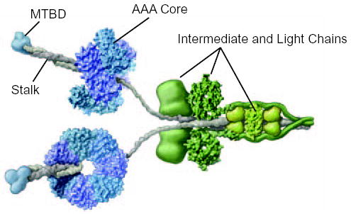
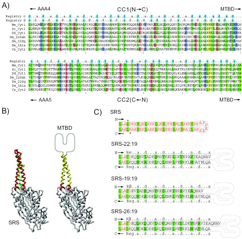
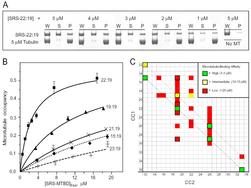
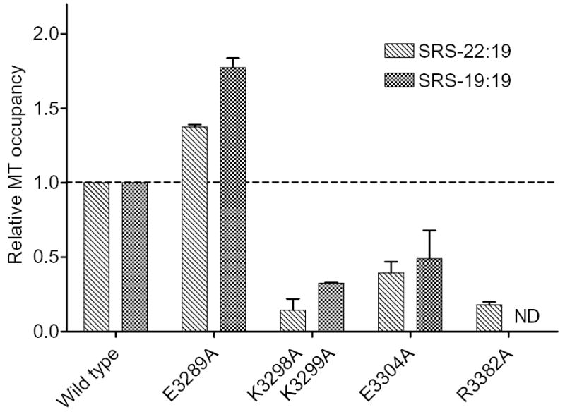
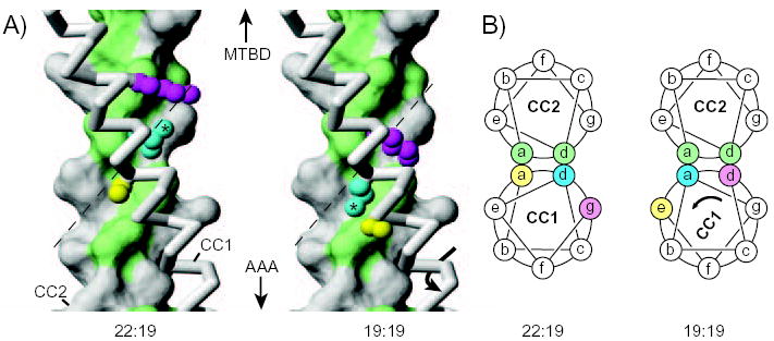
The microtubule-binding domain (MTBD) of dynein is separated from the AAA (ATPase with any other activity) core of the motor by an approximately 15-nm stalk that is predicted to consist of an antiparallel coiled coil. However, the structure of this coiled coil and the mechanism it uses to mediate communication between the MTBD and ATP-binding core are unknown. Here, we sought to identify the optimal alignment between the hydrophobic heptad repeats in the two strands of the stalk coiled coil. To do this, we fused the MTBD of mouse cytoplasmic dynein, together with 12-36 residues of its stalk, onto a stable coiled-coil base provided by Thermus thermophilus seryl-tRNA synthetase and tested these chimeric constructs for microtubule binding in vitro. The results identified one alignment that yielded a protein displaying high affinity for microtubules (2.2 microM). The effects of mutations applied to the MTBD of this construct paralleled those previously reported (Koonce, M. P., and Tikhonenko, I. (2000) Mol. Biol. Cell 11, 523-529) for an intact dynein motor unit in the absence of ATP, suggesting that it resembles the tight binding state of native intact dynein. All other alignments showed at least 10-fold lower affinity for microtubules with the exception of one, which had an intermediate affinity. Based on these results and on amino acid sequence analysis, we hypothesize that dynein utilizes small amounts of sliding displacement between the two strands of its coiled-coil stalk as a means of communication between the AAA core of the motor and the MTBD during the mechanochemical cycle.


Figures

FIG. 1
FIG. 1. Molecular structure of intact cytoplasmic dynein
The cytoplasmic dynein motor is a dimer containing two identical heavy chain subunits of Mm ~520 kDa. The core of the motor, formed by the C-terminal two-thirds of the heavy chain, comprises a ring of six AAA ATPase domains, depicted here in blue and purple. The microtubule-binding domain (MTBD) (blue) protrudes from the AAA core on a coiled coil stalk (grey). The attachment of cargo to the dynein motor involves light and intermediate chain subunits (green) that are associated with the N-terminal third of the heavy chain. Adapted from (2) with permission.
FIG. 2
FIG. 2. Design and construction of chimeric SRS-MTBD coiled coil
A, Sequence alignment of CC1 and CC2 region of dynein heavy chains. Species used are: mouse (Mm), Saccharomyces cerevisiae (Sc), Dictyostelium discoideum (Dd), Drosophila melanogaster (Dm), and Caenorhabditis elegans (Ce). Dynein isoforms used are: cytoplasmic (Cyt1), intra-flagellar transport (Cyt2), axonemal outer arm (22Sab and 22Sg), and axonemal inner arm (1A1). The sequences of CC2 are written in reverse order so that the ends of CC1 and CC2 adjoining the MTBD are both situated on the same (right) side of the alignment. In the mouse sequence (top line) the alignment shows CC1 residues from Leu3192 to His3301 and CC2 residues from Trp3393 to Ala3502. Positions in the alignment are shaded colours to indicate amino-acid type where the similarity is greater than 70%. The conserved residues Pro3285 and Pro3409 are highlighted yellow. Heptad repeats are indicated above the alignment: a, first position of heptad; d, fourth position of heptad. These heptad markers are shaded cyan at positions where there is >50% consensus of similar hydrophobic amino acids. The amino acids Ile, Leu, Val, Ala and Met are considered similar hydrophobic residues. B, Backbone trace of the SRS molecule used in SRS-MTBD constructs (PDB: 1SRY). Side chains of the amino acids forming the heptad repeat in the SRS coiled coil (left) are shown in green. In the SRS-MTBD cartoon (right), residues belonging to the SRS coiled-coil base are indicated in red and residues replaced by dynein are indicated in yellow. The MTBD located at the tip of the stalk is indicated schematically. C, Diagram showing the amino acid sequence of the coiled-coil stalk in native SRS and in 3 chimeric SRS-MTBD constructs. Asterisks adjacent to the SRS sequence indicate residues Leu33 and Arg93 that become mutated to Val and Lys, respectively, in order to generate SalI and HindIII restriction sites. The SRS-derived amino acids are indicated in red. Rα and Rβ α and β registries of CC1; Reg, registry of CC2. Amino acids comprising the “a” and “d” positions of the heptad repeats are highlighted in green. The conserved residues Pro3285 and Pro3409 are in magenta. The structure of the dynein MTBD is represented in cartoon form.
FIG. 3
FIG. 3. Binding of chimeric SRS-MTBD constructs to microtubules
A, Polyacrylamide electrophoresis gels showing the binding of different concentrations of SRS-22:19 to microtubules. Binding was assayed by co-sedimentation after incubation with a suspension of 5 μM microtubules and the indicated concentrations of SRS-22:19 for 15 min at room temperature. See Methods for assay details. W, uncentrifuged sample, S, supernatant; P, pellet. A parallel sample of SRS-22:19 incubated and centrifuged with no microtubules was used as a blank (typically 1–2%). Recovery of SRS-MTBD and tubulin averaged (94 ± 10)%. B, Microtubule binding affinity of SRS-MTBD constructs with a fixed CC2 length of 19 amino acids and different lengths of CC1. Assays and gel electrophoresis were performed essentially as in Fig. 3A. Error bars indicate standard error of 2 replicate gels of the same microtubule-binding assay. Averages of affinity data from multiple independent preparations are given in text. C, Affinity of microtubule binding by SRS-MTBD constructs with different lengths of CC1 and CC2. The affinity of 33 constructs with CC1/CC2 lengths ranging from 12 to 36 amino acids was classified as high, medium or low by comparing the fraction of SRS-MTBD bound to microtubules in side-by-side assays with one or more of the constructs whose affinity had been assayed in detail as shown in Fig 3B. Assays were performed with 3 μM and 10 μM SRS-MTBD with 5 μM microtubules. The shortest constructs, SRS-12:12 and SRS-15:12, were less stable than the others and assays involving them have been corrected for presence of a non-binding aggregated form. Diagonal lines indicate constructs related to SRS-22:19 (solid black) and SRS-19:19 (dashed black). Cells with thick black border indicate constructs for which Rs has been determined (Supplemental data).
FIG. 4
FIG. 4. Effect of site-directed MTBD mutations on microtubule binding by SRS-MTBD constructs
The microtubule occupancy of SRS-22:19 and SRS-19:19 constructs containing the indicated MTBD mutations is shown relative to that of corresponding constructs containing the wild-type MTBD. Microtubule occupancy was determined in side-by-side assays of mutant and wild-type constructs containing 3 μM SRS-22:19 or 10 μM SRS-19:19 with 5 μM microtubules. Bar heights show mean of 2 independent preparations for each mutated SRS-MTBD construct. Error bars show standard errors. ND, no data.
FIG. 5
FIG. 5. Structural models of the dynein stalk
A, Models of a section of the dynein stalk in configurations expected to correspond to those occurring in SRS-22:19 or SRS19:19. CC2 is shown in surface representation with hydrophobic residues (Val, Ile, Leu, Ala, Met, Tyr) in the coiled-coil core (“a” and “d” positions) coloured green. CC1 is shown in backbone representation (grey), with side chains included for Ser3224 (yellow), Leu3227 (cyan; with asterisk) and Lys3230 (magenta). In passing from the configuration of SRS-22:19 to that of SRS-19:19, the side chain of Leu3227 shifts from packing against one side of CC2 (left) to packing against the other side (right), potentially by following the hydrophobic-lined groove, formed by Ile3459, Leu3463, Val 3466 and Val 3470, in the surface of CC2 (dashed line). Similar hydrophobic grooves in the core interface of CC2 occur in most other heptads along the length of the stalk coiled coil (Supplemental Data). B, Cartoons depicting transverse sections through the coiled-coil stalk in the region shown in Fig. 5A. Residues in CC1 are coloured as in Fig. 5A to illustrate that Leu3227 (cyan) shifts from an “a” heptad position to a “d” position in going between the 22:19 and 19:19 configurations (see also Fig. 2A). Part A of this figure was prepared with the program MOLMOL (28).

References

    1. Gibbons IR. Cell Motil Cytoskeleton. 1995;32:136–144. - PubMed
    1. Vale RD. Cell. 2003;112:467–480. - PubMed
    1. Vallee RB, Williams JC, Varma D, Barnhart LE. J Neurobiol. 2004;58:189–200. - PubMed
    1. Porter ME, Sale WS. J Cell Biol. 2000;151:F37–F42. - PMC - PubMed
    1. Kamiya R. Int Rev Cytol. 2002;219:115–155. - PubMed

Publication types