

doi: 10.4103/ijmr.IJMR_577_20.
Transmission electron microscopy imaging of SARS-CoV-2
Collaborators,
Affiliations
- PMID: 32362648
- PMCID: PMC7224615
- DOI: 10.4103/ijmr.IJMR_577_20
Item in Clipboard
Transmission electron microscopy imaging of SARS-CoV-2
Indian J Med Res.
2020.
No abstract available
Conflict of interest statement
None
Figures
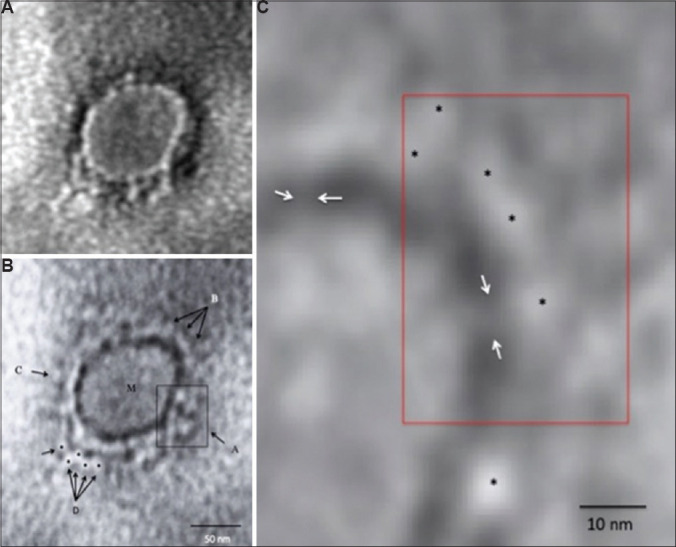
Transmission electron microscopy imaging of COVID-19. (A) A representative negative-stained COVID-19 particle showing morphodiagnostic features of family Coronaviridae. (B) Defocussed image of the same particle resolving the virus envelope glycoprotein morphology in finer details. The boxed area A shows a tetramer-like aggregate of four distinct peplomers, arrows shown by B show a more orthodox morphology of coronavirus surface projections. M indicates the matrix of the virus particle. C shows a distinct 'peplomer head' with negative stain silhouette. The area D is interesting as possible linear projections could be imaged. Five distinct peplomers could be imaged as shown by the arrows. (C) A highly magnified processed image for pixel corrections shows a distinct evidence of direct 'stalk' connecting the peplomer to the virion surface. The peplomers are shown with asterisk and the stalk with an arrow. Magnification bars are built into the micrographs.
References
-
- Gorbalenya AE, Baker SC, Baric RS, de Groot RJ, Drosten C, Gulyaeva AA, et al. The species severe acute respiratory syndrome-related coronavirus: classifying 2019-nCoV and naming it SARS-CoV-2. Nat Microbiol. 2020. https://doiorg/101038/s41564-020-0695-z . - PMC - PubMed
-
- Brenner S, Horne RW. A negative staining method for high resolution electron microscopy of viruses. Biochim Biophys Acta. 1959;34:103–10. - PubMed
-
- Madeley CR, Field AM. In: Virus morphology. 2nd ed. Field AME, editor. London: Churchill Livingstone; 1988.
Publication types
MeSH terms
LinkOut - more resources
Full Text Sources
Other Literature Sources
Miscellaneous