. 2013 Aug 7;3(8):1225-39.
doi: 10.1534/g3.112.002881.

Genomes of Ashbya fungi isolated from insects reveal four mating-type loci, numerous translocations, lack of transposons, and distinct gene duplications

Affiliations

Genomes of Ashbya fungi isolated from insects reveal four mating-type loci, numerous translocations, lack of transposons, and distinct gene duplications

Fred S Dietrich et al. G3 (Bethesda). .

Abstract

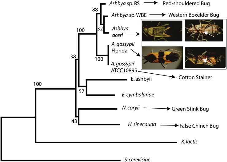
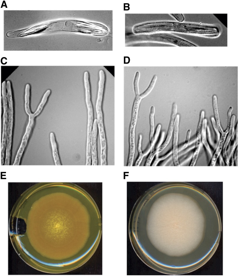
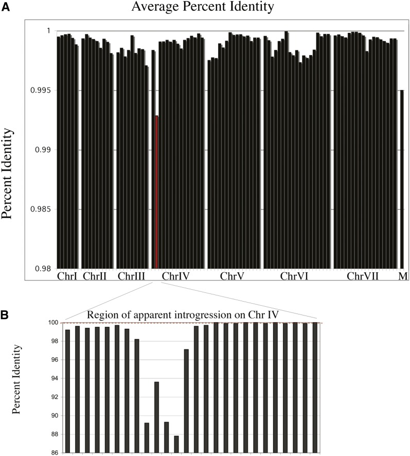
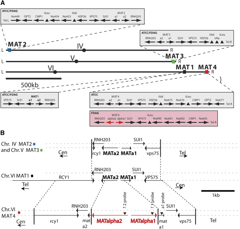
The filamentous fungus Ashbya gossypii is a cotton pathogen transmitted by insects. It is readily grown and manipulated in the laboratory and is commercially exploited as a natural overproducer of vitamin B2. Our previous genome analysis of A. gossypii isolate ATCC10895, collected in Trinidad nearly 100 years ago, revealed extensive synteny with the Saccharomyces cerevisiae genome, leading us to use it as a model organism to understand the evolution of filamentous growth. To further develop Ashbya as a model system, we have investigated the ecological niche of A. gossypii and isolated additional strains and a sibling species, both useful in comparative analysis. We isolated fungi morphologically similar to A. gossypii from different plant-feeding insects of the suborder Heteroptera, generated a phylogenetic tree based on rDNA-ITS sequences, and performed high coverage short read sequencing with one A. gossypii isolate from Florida, a new species, Ashbya aceri, isolated in North Carolina, and a genetically marked derivative of ATCC10895 intensively used for functional studies. In contrast to S. cerevisiae, all strains carry four not three mating type loci, adding a new puzzle in the evolution of Ashbya species. Another surprise was the genome identity of 99.9% between the Florida strain and ATCC10895, isolated in Trinidad. The A. aceri and A. gossypii genomes show conserved gene orders rearranged by eight translocations, 90% overall sequence identity, and fewer tandem duplications in the A. aceri genome. Both species lack transposable elements. Finally, our work identifies plant-feeding insects of the suborder Heteroptera as the most likely natural reservoir of Ashbya, and that infection of cotton and other plants may be incidental to the growth of the fungus in its insect host.

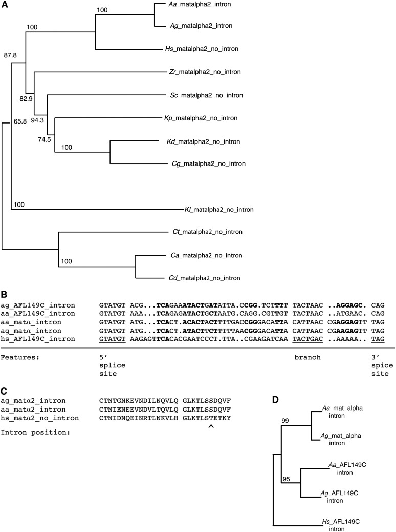
Keywords: fungal ecology; intron evolution; mating type; tandem duplications.


Figures

Figure 1
Figure 1
Fungi most closely related to A. gossypii. Neighbor joining phylogenetic tree of the known and newly isolated Nematosporaceae based on ITS sequences, along with Kluyveromyces lactis, and with S. cerevisiae as an outgroup. Ashbya sp. RS isolated from red-shouldered bugs and Ashbya sp. western boxelder (WBE) isolated from western boxelder bugs are as-yet uncharacterized beyond ITS sequencing. A. aceri is a fungus isolated from an eastern box elder bug shown in the insert. Its complete genome sequence was determined during this work. A. gossypii Florida isolate was isolated from a large milkweed bug shown in the insert. Its complete genome sequence also was determined during this work. The white arrows in the insect images indicate the probosci through which these insects feed, and through which the fungus is transmitted between the plant and the insect. A. gossypii has been described before to be spread by the cotton stainer (Ashby and Nowell 1926; Frazer 1944). A. gossypii ATCC10895 refers to the reference strain for all Ashbya species the genome of which was resequenced for the comparative analyses presented in this study This A. gossypii reference strain from the American Type Culture Collection was isolated from diseased cotton (Ashby 1916) and most likely originated from the US Agricultural Research Service strain collection (NRRL Y-1056), where it was obtained from William J. Robbins, who reported obtaining it from the Centraalbureau Voor Schimmelcultures (CBS) (Robbins and Schmidt 1939), where A. gossypii had been deposited by S. F. Ashby in 1926 (CBS 109.26), and possibly the same strain was deposited by Alexandre Guilliermond in 1928 (CBS 117.28). Holleya sinecada has been reported to be spread by the False Chinch bug (Burgess and McKenzie 1991), and Nematospora coryli by the Green stink bug (Clarke and Wilde 1970). Specific insect species have not been associated with Eremothecium cymbalariae or Eremothecium ashbyii. It has been suggested that all of the fungi of the family Nematosporaceae are spread by heteropterous insects (Batra 1973). GenBank accession numbers for the ITS sequences of the other Nematosporaceae are U09326.1 for Nematospora coryli, FJ422506.1 for Holleya sinecauda, AY046219.1 for Eremothecium cymbalariae, AB478315.1 for Eremothecium ashbyii, AJ229069.1 for the yeast Kluyveromyces lactis, and NC_001144 for Saccharomyces cerevisiae. The available ITS sequence data cannot well resolve the structure of the tree at the base of the Nematosporaceae clade.
Figure 2
Figure 2
Growth of A. gossypii insect isolate 1 strain and A. aceri (insect isolate 38). (A) Ascus from insect isolate 1. (B) Ascus from insect isolate 38. (C) Hyphal tip branching of insect isolate 1. (D) Hyphal tip branching of insect isolate 38. (E) Hyphal mat formed by insect isolate 1. (F) Hyphal mat formed by insect isolate 38. No aerial mycelial growth was observed.
Figure 3
Figure 3
Synteny between orthologous chromosomal regions of A. gossypii and S. cerevisiae. The yellow and red rectangles represent ORFs 267−294 of the right arm of chromosome 7 of A. gossypii ATCC10895 and insect isolate 1, respectively. The dark gray and light gray rectangles represent S. cerevisiae ORFs from the right arm of chromosome XV (above) and the left arm of chromosome XII (below), which are syntenic to the A. gossypii ORFs. Open triangles show transcription directions and filled arrow heads mark ORFs with intron. Open squares are tRNA genes and closed squares small nuclear RNA genes. The gene order is conserved between the two A. gossypii strains and also the lengths of the ORFs (number of codons) and the sizes of the inter-ORF regions (number of base pairs). The synteny with S. cerevisiae is divided between two chromosomal regions. At the time of the S. cerevisiae genome duplication both regions showed complete synteny to the A. gossypii gene order. During evolution many of the duplicated genes lost one copy seen as ORF-free regions in this synteny map. The synteny map also reveals six cases (five ORFs and one tRNA gene) where both copies of the duplication are retained. To distinguish these duplications from tandem duplications the term twin genes was coined (Dietrich et al. 2004).
Figure 4
Figure 4
Blocks of sequence conservation of up to 450 genes between ATCC10895 and insect isolate 1. (A) Distribution of sequence identity across the genomes was averaged over 100-kb intervals reveals that some regions are more similar, and some more diverged. On chromosome V the region from approximately 501,000 to 1,270,000, spanning 410 protein coding genes is 99.96% identical between these strains. On chromosome VI the region from approximately 700,000 to 1,478,000, spanning 434 protein coding genes, is 99.80% identical between these strains. The mitochondrial genome labeled “M” is more diverged than the nuclear genome. The telomeric regions of chromosomes V, VI, and VII show more sequence divergence, particularly rearrangements in repetitive elements, than the genome overall and are not shown in this figure. The nuclear genomes are on average 99.9% identical, excluding the telomeric regions. (B) A syntenic region of 5450 bases of significantly lower homology, approximately 92% identity, between A. gossypii strains ATCC10895 and insect isolate 1 is found on chromosome 4 (red bar in A), with boundaries from 179,139 to 184,589 bases in ATCC10895. Percent identity was averaged over windows of 1 kb. Of the 439 SNPs in the introgression region, 139 are in inter-ORF regions, which have an average identity of 92.6%. The remaining 300 SNPs, 186 synonymous and 97 nonsynonymous, fall in the four open reading frames of this region, ADL294C, ADL295W, ADL296C, and ADL297W, which have an average identity of 91.6%. Interestingly, one of the genes, ADL296C, encodes the enzyme GTP cyclohydrolase, the first step in riboflavin biosynthesis. Although the introgressed regions are 92% identical to each other, they are both approximately equally diverged from A. aceri at only 78% identity each, suggesting the source of the introgression is not A. aceri, but another Ashbya species more closely related to A. gossypii.
Figure 5
Figure 5
A. gossypii mating type regions of ATCC10895 and the insect isolate 1 strain. (A) Overall organization of the four mating type loci on chromosomes IV, V, and VI. The three chromosomes are shown in the orientation as annotated. Circles mark the centromere locations; colored squares mark the locations of the mating type loci MAT1 to MAT4. The enlarged sections show the genetic map of these regions in both strains. No differences were found except for the MAT4 locus at the right telomere of chromosome 6 that carries α1/α2 information in the Florida isolate and a1/a2 information in ATCC10895. Interestingly, the order of genes distal to MAT1 and MAT4 is identical in ATCC10895. It is therefore very likely that the MAT4 locus of ATCC10895 originally carried α1/α2 genes, like the Florida isolate, which were replaced with a1/a2 genes by a gene conversion event with the left telomere of chromosome 4 initiated by a break in the homology region around RNH203 proximal to MAT4. Table S2 presents the nomenclature of genes associated with the four MAT loci. (B) Fine structure of the four MAT loci of the Florida isolate and ATCC10895 before the gene conversion at MAT4. All gene names refer to the S. cerevisiae homologs, except for a2, which is a homolog of the K. lactis MATa2 gene (Astrom et al. 2000). The telomeric loci on chromosomes IV and V are flanked by partial copies of the RCY1 and VPS75 genes, marked in lower case. Vertical bars and dotted lines indicate the junctions of homology at the mating type loci, the centromeric and telomeric ends being marked by “Cen” and “Tel.” Only the nontelomeric MAT1 locus is flanked by intact RCY1 and VPS75 genes, suggesting that this locus on chromosome VI is the active mating type locus, with ATCC10895 and the Florida isolate 1 being MATa. The orientation shown is opposite of that in part A. The chromosome VI telomeric MAT4 locus in the Florida strain carries MATα2 and MATα1 genes inserted into remnants of MATa2 and MATa1 genes, indicated in lower case. The locus is somewhat larger, containing more sequence from the still truncated RCY1 and VPS75 genes. MATα specific sequences are shown in red. The sequence arrangements at the MAT loci were confirmed by DNA hybridizations using synthetic oligonucleotides with homology to the positions indicated by arrow heads (data not shown).
Figure 6
Figure 6
An unusual intron in Ashbya MATα2. (A) A neighbor joining phylogenetic tree of the MATα2 protein. Numbers refer to bootstrap values. Species are A. aceri (Aa), A. gossypii (Ag), H. sinecauda (Hs), S. cerevisiae (Sc), Kluyveromyces (Vanderwaltozyma) polyspora (Kp) (Scannell et al. 2007), Kluyveromyces delphensis (Kd) (Wong et al. 2003), Candida glabrata, (Cg) (Dujon et al. 2004), Zygosaccharomyces rouxi (Zr) (Souciet et al. 2009), Kluyveromyces lactis (Kl) (Dujon et al. 2004), Candida thermotolerans (Ct) (Souciet et al. 2009), Candida albicans (Ca) (Hull and Johnson 1999), and Candida dubliniensis (Cd). (B) Alignment of the introns of A. gossypii gene AFL149C with the homologous introns from H. sinecauda and A. aceri, and the introns from the MATα2 gene of A. gossypii and A. aceri. An intron at this position is found in no other MATα2 genes currently available in GenBank. The 5′ splice site, branch point, and 3′ splice site are marked. Conserved sequence within the intron is marked in bold. (C) A partial alignment of the Ashbya and H. sinecauda MATα2 proteins is shown with the position of the intron marked. The intron is outside the conserved homeobox domain. The alignment suggests no sequences have been gained or lost at the site of this intron. (D) A phylogenetic tree of the intron sequences of MATα2 and AFL149C.
Figure 7
Figure 7
Unique case of an intron with two 3′ splice sites in Ashbya. (A) The intron in A. gossypii ADR221C is shown to scale with the two branch points and two 3′ splice sites. (B) The sequence of the A. gossypii ADR221C intron, total size 105 nucleotides for the longer form. The 5′ Splice site, two branch points, and two 3′ splice sites are shown in upper case, with the translation below. (C) A. aceri ADR221C intron, with a total size 110 nucleotides for the longer form.

References

    1. Altmann-Jöhl R., Philippsen P., 1996. AgTHR4, a new selection marker for transformation of the filamentous fungus Ashbya gossypii, maps in a four-gene cluster that is conserved between A. gossypii and Saccharomyces cerevisiae. Mol. Gen. Genet. 250: 69–80 - PubMed
    1. Altschul S. F., Madden T. L., Schaffer A. A., Zhang J., Zhang Z., et al. , 1997. Gapped BLAST and PSI-BLAST: a new generation of protein database search programs. Nucleic Acids Res. 25: 3389–3402 - PMC - PubMed
    1. Ashby S. F., 1916. Annual Report of the Microbiologist, 1915–1916. Annual report of the Department of Agriculture, Jamaica, pp. 29–31
    1. Ashby S. F., Nowell W., 1926. The fungi of Stigmatomycosis. Ann. Bot. (Lond.) 40: 69–83
    1. Astrom S. U., Kegel A., Sjostrand J. O., Rine J., 2000. Kluyveromyces lactis Sir2p regulates cation sensitivity and maintains a specialized chromatin structure at the cryptic alpha-locus. Genetics 156: 81–91 - PMC - PubMed

Publication types

Associated data

LinkOut - more resources