. 2006 Jul 11:7:21.
doi: 10.1186/1471-2199-7-21.

Multiple erythroid isoforms of human long-chain acyl-CoA synthetases are produced by switch of the fatty acid gate domains

Affiliations

Multiple erythroid isoforms of human long-chain acyl-CoA synthetases are produced by switch of the fatty acid gate domains

Eric Soupene et al. BMC Mol Biol. .

Abstract

Background: The formation of acyl-CoA by the action of acyl-CoA synthetases plays a crucial role in membrane lipid turnover, including the plasma membrane of erythrocytes. In human, five Acyl-CoA Synthetase Long-chain (ACSL) genes have been identified with as many as 3 different transcript variants for each.

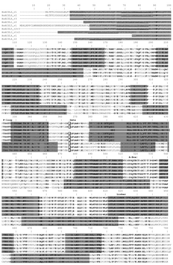
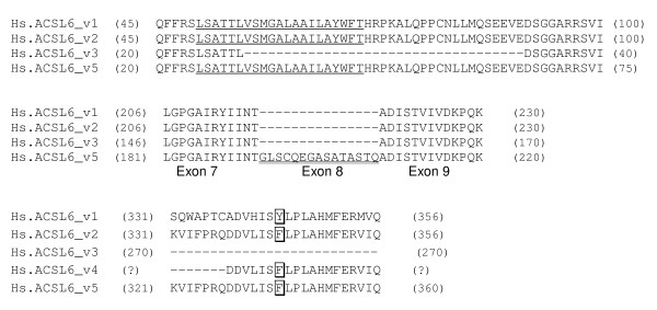
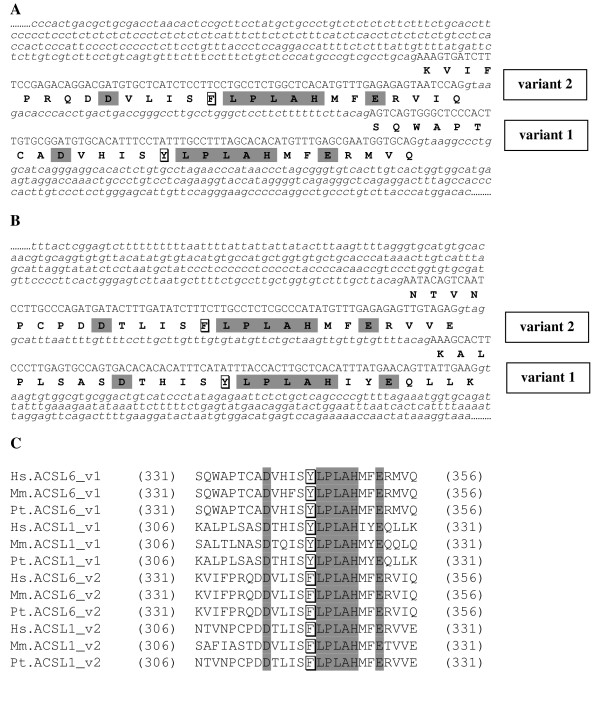
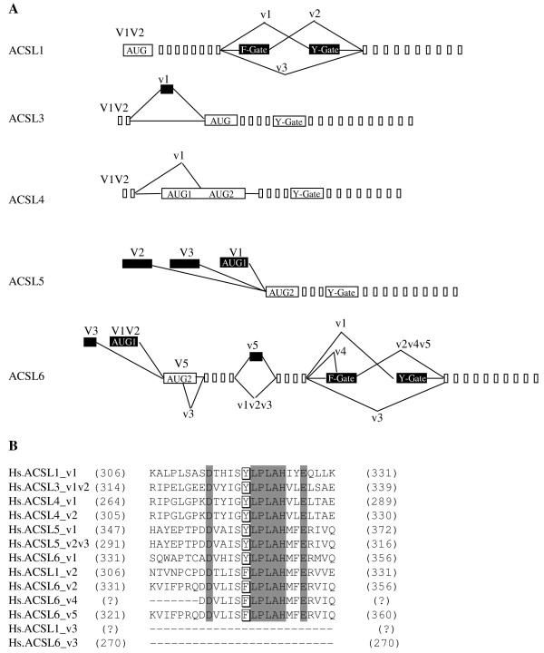
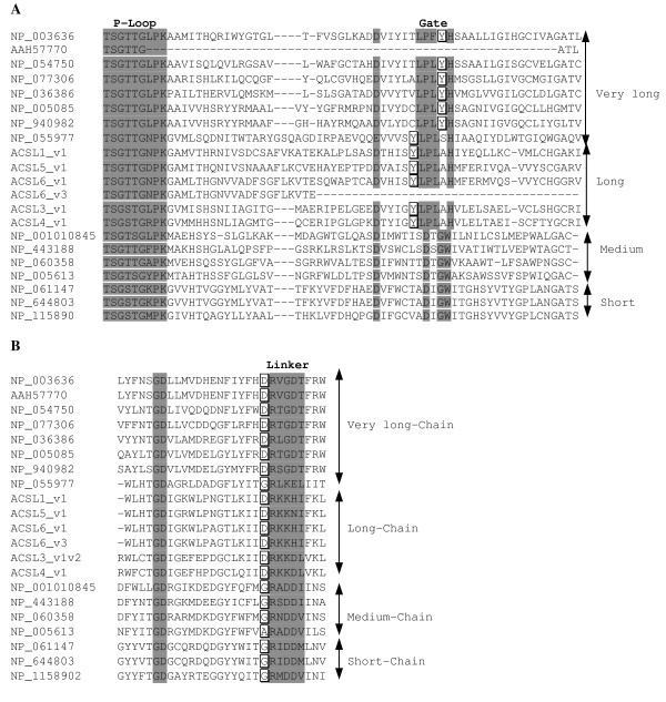
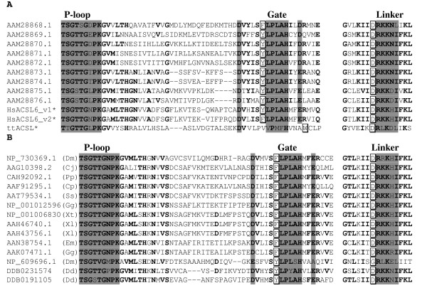
Results: Acyl-CoA Synthetase Long-chain member 6 (ACSL6) is responsible for activation of long-chain fatty acids in erythrocytes. Two additional transcript variants were also isolated from brain and testis. We report the expression in reticulocytes of two new variants and of the one isolated from brain. All three represented different spliced variants of a mutually exclusive exon pair. They encode a slightly different short motif which contains a conserved structural domain, the fatty acid Gate domain. The motifs differ in the presence of either the aromatic residue phenylalanine (Phe) or tyrosine (Tyr). Based on homology, two new isoforms for the closely related ACSL1 were predicted and characterized. One represented a switch of the Phe- to the Tyr-Gate domain motif, the other resulted from the exclusion of both. Swapping of this motif also appears to be common in all mammalian ACSL member 1 and 6 homologs.

Conclusion: We propose that a Phe to Tyr substitution or deletion of the Gate domain, is the structural reason for the conserved alternative splicing that affects these motifs. Our findings support our hypothesis that this region is structurally important to define the activity of these enzymes.


Figures

Figure 1
Figure 1
Sequence alignment of the human acyl-CoA synthetases long-chain family members. The succession of clear-shaded blocks overlaid on the alignment represents the exon organization of the respective mRNA. Conserved residues are in bold. Position of the P-loop, Gate, A-box and Linker motifs is indicated. Y and F residues of the two alternative Gate domains are boxed and indicated with a star. Transmembrane spanning segments predicted with the highest score by TopoPrediction, SOSUI and TMHMM are underlined. For clarity, ACSL3 variant 1 and 2, and ACSL5 variant 2 and 3, which encode the same isoform, are annotated as ACSL3_v1v2 and ACSL5_v2v3, respectively. Alignment was performed with MUTALIN [14]. Protein length (residues): ACSL1_v1, 698; ACSL1_v2, 698; ACS3_v1v2, 720; ACSL4_v1, 670; ACSL4_v2, 711; ACSL5_v1, 739; ACSL5_v2v3, 683; ACSL6_v1, 722; ACSL6_v2, 722; ACSL6_v3, 622. GenBank accessions numbers are given in Table 2.
Figure 2
Figure 2
Details of the sequence differences of the five human ACSL6 isoforms. The Y and F residues of the Gate domains are boxed. Position of the first and last residues shown is indicated on the left and right, respectively. Note that only the Gate-domain region of ACSL6_v4 is known. A predicted transmembrane spanning segment at the amino-terminus is shown underlined. The inserted sequence in between Exon 7 and Exon 9 of variant 5, annotated as Exon 8, is shown double-underlined.
Figure 3
Figure 3
Detection of ACSL6 isoforms in E. coli (A) and in human K562 cells (B). A: vector (1, 2, 3); ACSL6_v2 (4, 5, 6); ACSL6_v3 (7, 8, 9). Crude extract (1, 4, 7); Soluble fraction (2, 5, 8); Membrane fraction (3, 6, 9). Detection performed with Pierce INDIA HIS-antibody. Protein ladder standard is shown on the right (top to bottom: 250-150-100-75 kDa). Proteins carried a hexahistidine-tag at their amino-terminus (see Methods). B: Total protein extract of K562 cells. Detection performed with a peptide raised antibody against ACSL6.
Figure 4
Figure 4
Genomic and amino acid sequences of the gate-domain region of human ACSL6 and ACSL1. A and B: Sequence of exons and introns of human ACSL6 (A) and ACSL1 (B) are represented by capitalized and small italics letters, respectively. The F and Y residues of the Gate domain of variant 2 and 1, respectively, are boxed. C: Amino acids alignment of the Gate-domain motifs for known and predicted spliced variants. Highly conserved residues are shaded. Position of the first and last residues shown is indicated on the left and right, respectively. Abbreviations are: Hs, Homo sapiens; Ms, Mus musculus; Pt, Pan troglodytes.
Figure 5
Figure 5
Human ACSL transcript variants (A) and gate domains (B). A: Schematic representation of the different known transcript variants. Exons are represented by empty boxes and alternative exons by filled boxes. Translation initiator AUG and Y/F-Gate domain encoding exons are indicated. If known, a capitalized V indicates the first exon of each variant. Note that only the sequence of the Gate-domain region of ACSL1_v3 and ACSL6_v4 is known. See Results for details. B: Alignment of the Gate-domain motifs. The F and Y residues of the two Gate domains are boxed. Highly conserved residues are shaded. Note that more residues are conserved among human ACSL isoforms. Position of the first and last residues shown is indicated on the left and right, respectively. Note that only the Gate-domain region of ACSL1_v3 and ACSL6_v4 is known. For clarity, ACSL3 variant 1 and 2, and ACSL5 variant 2 and 3, which encode the same isoform, are annotated as ACSL3_v1v2 and ACSL5_v2v3, respectively.
Figure 6
Figure 6
Gate (A) and linker (B) domains of human acyl-CoA synthetase family members. The Y residue of the Gate domain for long and very-long-chain ACS is boxed. The D and G residues of the Linker domains are boxed. GenBank accession numbers for short, medium, long and very-long chain are indicated on the left. The Gate domain of BG1 is of the long-chain type but its Linker is of the short-medium type. Lipidosin is a soluble ACS very-long-chain isoform found in brain tissue and is known to have a broader substrate range than other very-long ACS [17, 18].
Figure 7
Figure 7
Conservation of the gate and linker motifs among ACSL homologs. A: Alignment of the nine Arabidopsis ACSL forms. Human ACSL6 spliced variant 1 and 2, and ACSL of Thermus thermophilus (ttACSL) indicated with an asterisk (*) are shown as references in the last three rows. B: Alignment of various forms of ACSL from different organisms. Abbreviations: Dm, Drosophila melanogaster (fly); Cj, Callithrix jacchus (marmoset); Pp, Pongo pygmaeus (orangutan); Cp, Cavia porcellus (guinea pig); Ss, Sus scrofa (pig); Gg, Gallus gallus (chicken); Xt, Xenopus tropicalis (frog); Xl, Xenopus laevis (frog); Em, Eleginops maclovinus (fish); Gg, Gobionotothen gibberifrons (fish); Dd, Dictyostelium discoideum (slime mould).

References

    1. Lands WE, Hart P. Metabolism Of Glycerolipids. Vi. Specificities Of Acyl Coenzyme A: Phospholipid Acyltransferases. J Biol Chem. 1965;240:1905–1911. - PubMed
    1. Malhotra KT, Malhotra K, Lubin BH, Kuypers FA. Identification and molecular characterization of acyl-CoA synthetase in human erythrocytes and erythroid precursors. Biochem J. 1999;344:135–143. doi: 10.1042/0264-6021:3440135. - DOI - PMC - PubMed
    1. Hisanaga Y, Ago H, Nakagawa N, Hamada K, Ida K, Yamamoto M, Hori T, Arii Y, Sugahara M, Kuramitsu S, Yokoyama S, Miyano M. Structural basis of the substrate-specific two-step catalysis of long chain fatty acyl-CoA synthetase dimer. J Biol Chem. 2004;279:31717–31726. doi: 10.1074/jbc.M400100200. - DOI - PubMed
    1. Weimar JD, DiRusso CC, Delio R, Black PN. Functional role of fatty acyl-coenzyme A synthetase in the transmembrane movement and activation of exogenous long-chain fatty acids. Amino acid residues within the ATP/AMP signature motif of Escherichia coli FadD are required for enzyme activity and fatty acid transport. J Biol Chem. 2002;277:29369–29376. doi: 10.1074/jbc.M107022200. - DOI - PubMed
    1. Lewin TM, Van Horn CG, Krisans SK, Coleman RA. Rat liver acyl-CoA synthetase 4 is a peripheral-membrane protein located in two distinct subcellular organelles, peroxisomes, and mitochondrial-associated membrane. Arch Biochem Biophys. 2002;404:263–270. doi: 10.1016/S0003-9861(02)00247-3. - DOI - PubMed

Publication types

MeSH terms

Associated data

LinkOut - more resources