. 2006 Apr 25;103(17):6724-9.
doi: 10.1073/pnas.0510644103. Epub 2006 Apr 14.

ADP-ribosylating and vacuolating cytotoxin of Mycoplasma pneumoniae represents unique virulence determinant among bacterial pathogens

Affiliations

ADP-ribosylating and vacuolating cytotoxin of Mycoplasma pneumoniae represents unique virulence determinant among bacterial pathogens

T R Kannan et al. Proc Natl Acad Sci U S A. .

Abstract

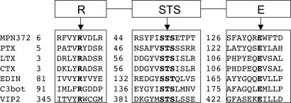
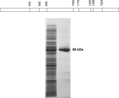
Unlike many bacterial pathogens, Mycoplasma pneumoniae is not known to produce classical toxins, and precisely how M. pneumoniae injures the respiratory epithelium has remained a mystery for >50 years. Here, we report the identification of a virulence factor (MPN372) possibly responsible for airway cellular damage and other sequelae associated with M. pneumoniae infections in humans. We show that M. pneumoniae MPN372 encodes a 68-kDa protein that possesses ADP-ribosyltransferase (ART) activity. Within its N terminus, MPN372 contains key amino acids associated with NAD binding and ADP-ribosylating activity, similar to pertussis toxin (PTX) S1 subunit (PTX-S1). Interestingly, MPN372 ADP ribosylates both identical and distinct mammalian proteins when compared with PTX-S1. Remarkably, MPN372 elicits extensive vacuolization and ultimate cell death of mammalian cells, including distinct and progressive patterns of cytopathology in tracheal rings in organ culture that had been previously ascribed to infection with WT virulent M. pneumoniae. We observed dramatic seroconversion to MPN372 in patients diagnosed with M. pneumoniae-associated pneumonia, indicating that this toxin is synthesized in vivo and possesses highly immunogenic epitopes.


Conflict of interest statement

Conflict of interest statement: No conflicts declared.

Figures

Fig. 1.
Fig. 1.
Alignment of conserved residues between MPN372 and other ARTs. Residues necessary for NAD-binding and catalysis are shown in bold face. PTX, B. pertussis pertussis toxin; LTX, E. coli heat-labile enterotoxin; CTX, cholera toxin; EDIN, Staphylococcus aureus epidermal cell differentiation inhibitor; C3bot, Clostridium botulinum C3 toxin; VIP2, Bacillus cereus vegetative insecticidal protein.
Fig. 2.
Fig. 2.
Expression and purification of CARDS TX protein. (Upper) Distribution of UGA codon within mpn372. The eight TGA codons within the coding region of CARDS TX were modified into TGG codons (at nucleotide positions shown in the schematic diagram) to express in E. coli. (Lower) CARDS TX gene was cloned in pET19b vector and expressed in E. coli BL21(DE3). Recombinant His-10-tagged protein was purified by using nickel affinity column chromatography and eluted by imidazole. Proteins were resolved in 4–15% gradient SDS/PAGE gel. Lane 1, overexpressed rCARDS TX in E. coli BL21(λDE3); lane 2, purified rCARDS TX.
Fig. 3.
Fig. 3.
CARDS TX-mediated ADP ribosylation of mammalian cell proteins. (a) ADP ribosylation of CHO cell-free extracts by rCARDS TX. CFEs were prepared from confluent CHO cell monolayers and assayed for ADP ribosylation. CFEs were incubated with and without rCARDS TX. The reaction mixture was precipitated with trichloroacetic acid (TCA), and proteins were resolved by gradient SDS/PAGE and transferred to nitrocellulose membrane for autoradiography as shown. Lanes: 1, CFE alone; 2, CFE + rCARDS TX; 3, CFE + rCARDS TX − DTT; 4, CFE + rCARDS TX − ATP; 5, CFE + rCARDS TX − GTP. (b) ADP ribosylation of HEp-2 cell proteins by rCARDS TX or PTX. HEp-2 cell monolayers were incubated with medium alone or in the presence of rCARDS TX or PTX (holotoxin). Cells were washed and incubated with fresh medium, and CFEs were prepared and assayed for ADP ribosylation. The reaction mixture was precipitated with trichloroacetic acid (TCA), and proteins were resolved by SDS/PAGE and transferred to nitrocellulose membrane for autoradiography. Lanes: 1, cells in medium alone followed by preparation of CFE and addition of rCARDS TX; 2, cells pretreated with rCARDS TX followed by preparation of CFE and addition of rCARDS TX; 3, cells pretreated with PTX followed by preparation of CFE and addition of rCARDS TX; 4, cells in medium alone followed by preparation of CFE and addition of PTX.
Fig. 4.
Fig. 4.
Effect of rCARDS TX on CHO cell morphology. Cells were grown to 60% monolayer confluence before addition of 10 μg of rCARDS TX for 16–40 h. Control CHO cells were treated with 10 μg of heat-inactivated rCARDS TX for 40 h. (Magnification: ×200.)
Fig. 5.
Fig. 5.
Effect of rCARDS TX on baboon tracheal epithelium. Baboon tracheal rings were incubated with 1.5, 5, or 10 μg of CARDS TX for 24–48 h in 5 ml of DMEM. Control baboon tracheal rings were treated with heat-inactivated CARDS TX for 48 h. (Magnification: ×200.)
Fig. 6.
Fig. 6.
ELISA-based screening of normal and M. pneumoniae-infected individuals for CARDS TX-reactive antibodies. Each well of ELISA plates was coated with rCARDS TX and reacted with patient sera, which were collected at the onset of disease (I) and 14 (II) and 28 (III) days later.

References

    1. Eaton M. D., Meikejohn G., Van Herick W. J. Exp. Med. 1944;79:649–667. - PMC - PubMed
    1. Couch R. B., Cate T. R., Chanock R. M. J. Am. Med. Assoc. 1964;187:442–447. - PubMed
    1. Feizi T., Taylor-Robinson D. Immunology. 1967;13:405–409. - PMC - PubMed
    1. Baseman J. B., Tully J. G. Emerg. Infect. Dis. 1997;3:21–32. - PMC - PubMed
    1. Baseman J. B., Reddy S. P., Dallo S. F. Am. J. Respir. Crit. Care Med. 1996;154:S137–S144. - PubMed

Publication types

MeSH terms