Observational Study
. 2021 Mar;27(3):458-466.
doi: 10.1016/j.cmi.2020.11.005. Epub 2020 Nov 13.

Neurologic manifestations associated with COVID-19: a multicentre registry

Collaborators, Affiliations
Observational Study

Neurologic manifestations associated with COVID-19: a multicentre registry

Elodie Meppiel et al. Clin Microbiol Infect. 2021 Mar.

Abstract

Objectives: To provide an overview of the spectrum, characteristics and outcomes of neurologic manifestations associated with severe acute respiratory syndrome coronavirus 2 (SARS-CoV-2) infection.

Methods: We conducted a single-centre retrospective study during the French coronavirus disease 2019 (COVID-19) epidemic in March-April 2020. All COVID-19 patients with de novo neurologic manifestations were eligible.

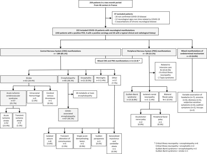
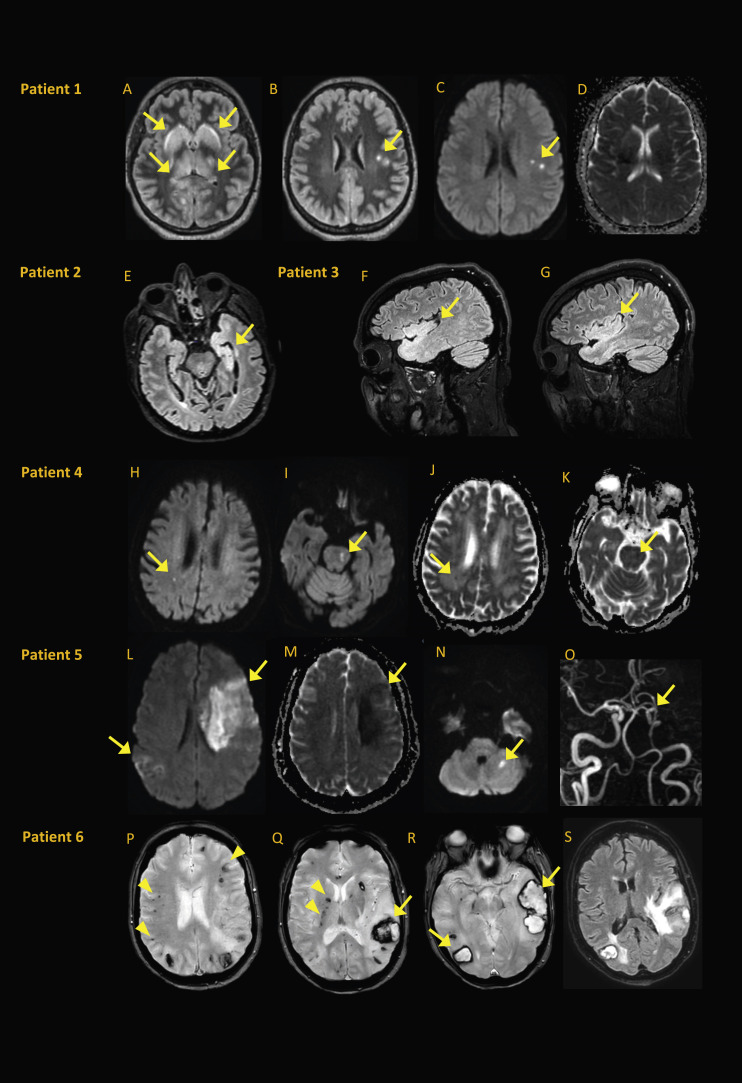
Results: We included 222 COVID-19 patients with neurologic manifestations from 46 centres in France. Median (interquartile range, IQR) age was 65 (53-72) years and 136 patients (61.3%) were male. COVID-19 was severe or critical in 102 patients (45.2%). The most common neurologic diseases were COVID-19-associated encephalopathy (67/222, 30.2%), acute ischaemic cerebrovascular syndrome (57/222, 25.7%), encephalitis (21/222, 9.5%) and Guillain-Barré syndrome (15/222, 6.8%). Neurologic manifestations appeared after the first COVID-19 symptoms with a median (IQR) delay of 6 (3-8) days in COVID-19-associated encephalopathy, 7 (5-10) days in encephalitis, 12 (7-18) days in acute ischaemic cerebrovascular syndrome and 18 (15-28) days in Guillain-Barré syndrome. Brain imaging was performed in 192 patients (86.5%), including 157 magnetic resonance imaging (70.7%). Among patients with acute ischaemic cerebrovascular syndrome, 13 (22.8%) of 57 had multiterritory ischaemic strokes, with large vessel thrombosis in 16 (28.1%) of 57. Brain magnetic resonance imaging of encephalitis patients showed heterogeneous acute nonvascular lesions in 14 (66.7%) of 21. Cerebrospinal fluid of 97 patients (43.7%) was analysed, with pleocytosis found in 18 patients (18.6%) and a positive SARS-CoV-2 PCR result in two patients with encephalitis. The median (IQR) follow-up was 24 (17-34) days with a high short-term mortality rate (28/222, 12.6%).

Conclusions: Clinical spectrum and outcomes of neurologic manifestations associated with SARS-CoV-2 infection were broad and heterogeneous, suggesting different underlying pathogenic processes.

Keywords: COVID-19; Nervous system; Neurologic manifestations; Registry; SARS-CoV-2.


Figures

Fig. 1
Fig. 1
Study population of coronavirus disease 2019 (COVID-19) patients with neurologic manifestations.
Fig. 2
Fig. 2
Brain MRI from patients with encephalitis or atypical strokes. (A–D) Patient 1∗, a 56-year-old woman with encephalitis, experienced headache, confusion, facial palsy, ophthalmoparesis, refractory status epilepticus and pleocytosis. SARS-CoV-2 PCR results were positive in respiratory sample but negative in CSF. Bilateral basal ganglia and thalami exhibited FLAIR hyperintensity (A), with small subcortical white matter FLAIR hyperintensities (B) visible in diffusion (C) with normal ADC map (D). (E) Patient 2, a 58-year-old man with encephalitis, was found to be SARS-CoV-2 PCR positive in nasopharyngeal swab sample and negative in CSF sample. Pleocytosis and left mesiotemporal and temporopolar hyperintensity were evident on axial FLAIR (E). (F, G) Patient 3, a 49-year-old man with encephalitis, experienced psychomotor agitation and inattention after withdrawal of sedation. SARS-CoV-2 PCR was positive in nasopharyngeal swab sample and negative in CSF. Bilateral temporal and insular hyperintensities were evident on sagittal FLAIR (F, G). (H–K) Patient 4, a 76-year-old man with encephalopathy, had altered mental status 14 days after severe respiratory symptoms. SARS-CoV-2 PCR results were positive in nasopharyngeal sample and negative in CSF; no pleocytosis was noted. Small diffusion hyperintensities were evident in right periventricular white matter (H) and left side of pons (I). Both lesions had decreased ADC (J, K) consistent with small acute ischaemic lesions that did not explain encephalopathy. (L–O) Patient 5, a 60-year-old woman with acute ischaemic stroke, experienced sudden right haemiparesis 11 days after severe respiratory symptoms. SARS-CoV-2 PCR results were positive in nasopharyngeal sample; assessment was negative for stroke and vascular risk factors. Diffusion hyperintensities were evident in left frontal and right parietal areas (L) and in left cerebellum (N), with decreased ADC (M) and left middle cerebral artery occlusion on time-of-flight magnetic resonance angiography (O). (P–S) Patient 6, a 60-year-old woman with multiple intracranial haemorrhages, experienced sudden right haemiparesis and aphasia after withdrawal of sedation. SARS-CoV-2 PCR results were positive in nasopharyngeal sample. Multiple hypointensities on axial gradient echo T2-weighted images were consistent with cortical microhemorrhages (arrowheads in P), deep microhemorrhages (arrowheads in Q) and haematoma in left parietal lobe (arrow in Q) occipital and temporal lobes (arrows in R) with perilesional oedema on axial FLAIR (S). ADC, apparent diffusion coefficient; CSF, cerebrospinal fluid; FLAIR, fluid-attenuated inversion recovery; MRI, magnetic resonance imaging; SARS-CoV-2, severe acute respiratory syndrome coronavirus 2. ∗Image courtesy of Dr F.Bruneel, Intensive Care Unit, Versailles Hospital; Image courtesy of Dr L.Dubuc, Neurology, Saint-Lo Hospital.

References

    1. Chen N., Zhou M., Dong X., Qu J., Gong F., Han Y. Epidemiological and clinical characteristics of 99 cases of 2019 novel coronavirus pneumonia in Wuhan, China: a descriptive study. Lancet. 2020;395:507–513. - PMC - PubMed
    1. Goyal P., Choi J.J., Pinheiro L.C., Schenck E.J., Chen R., Jabri A. Clinical characteristics of covid-19 in New York City. N Engl J Med. 2020 - PMC - PubMed
    1. Mao L., Jin H., Wang M., Hu Y., Chen S., He Q. Neurologic manifestations of hospitalized patients with coronavirus disease 2019 in Wuhan, China. JAMA Neurol. 2020 - PMC - PubMed
    1. Helms J., Kremer S., Merdji H., Clere-Jehl R., Schenck M., Kummerlen C. Neurologic features in severe SARS-CoV-2 infection. N Engl J Med. 2020 - PMC - PubMed
    1. Zubair A.S., McAlpine L.S., Gardin T., Farhadian S., Kuruvilla D.E., Spudich S. Neuropathogenesis and neurologic manifestations of the coronaviruses in the age of coronavirus disease 2019: a review. JAMA Neurol. 2020 - PMC - PubMed

Publication types