. 2007 Apr;19(4):1362-75.
doi: 10.1105/tpc.106.042911. Epub 2007 Apr 6.

Rice NON-YELLOW COLORING1 is involved in light-harvesting complex II and grana degradation during leaf senescence

Affiliations

Rice NON-YELLOW COLORING1 is involved in light-harvesting complex II and grana degradation during leaf senescence

Makoto Kusaba et al. Plant Cell. 2007 Apr.

Abstract

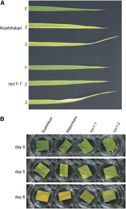
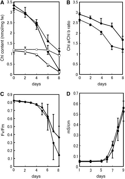
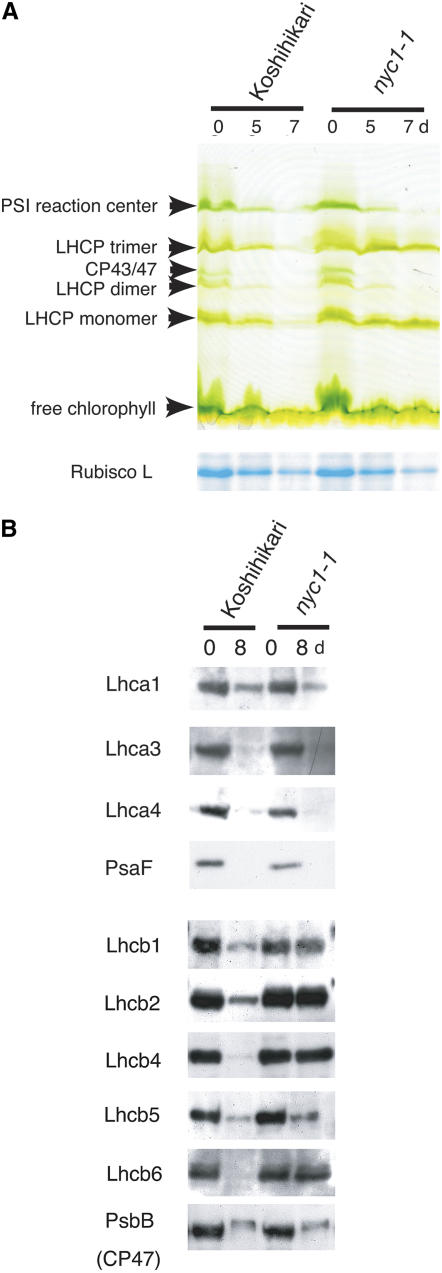
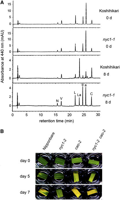
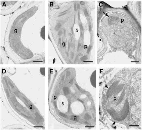
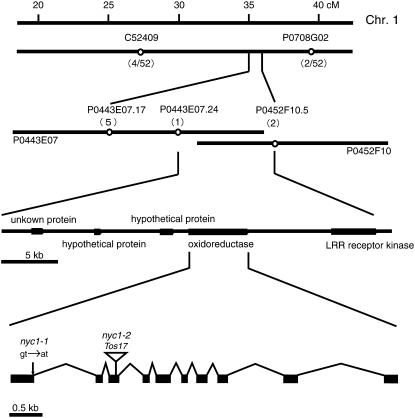
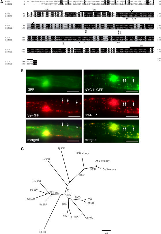
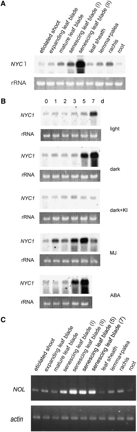
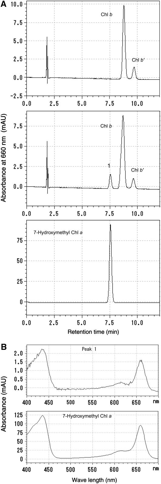
Chlorophyll degradation is an aspect of leaf senescence, which is an active process to salvage nutrients from old tissues. non-yellow coloring1 (nyc1) is a rice (Oryza sativa) stay-green mutant in which chlorophyll degradation during senescence is impaired. Pigment analysis revealed that degradation of not only chlorophylls but also light-harvesting complex II (LHCII)-bound carotenoids was repressed in nyc1, in which most LHCII isoforms were selectively retained during senescence. Ultrastructural analysis of nyc1 chloroplasts revealed that large and thick grana were present even in the late stage of senescence, suggesting that degradation of LHCII is required for the proper degeneration of thylakoid membranes. Map-based cloning of NYC1 revealed that it encodes a chloroplast-localized short-chain dehydrogenase/reductase (SDR) with three transmembrane domains. The predicted structure of the NYC1 protein and the phenotype of the nyc1 mutant suggest the possibility that NYC1 is a chlorophyll b reductase. Although we were unable to detect the chlorophyll b reductase activity of NYC1, NOL (for NYC1-like), a protein closely related to NYC1 in rice, showed chlorophyll b reductase activity in vitro. We suggest that NYC1 and NOL encode chlorophyll b reductases with divergent functions. Our data collectively suggest that the identified SDR protein NYC1 plays essential roles in the regulation of LHCII and thylakoid membrane degradation during senescence.


Figures

Figure 1.
Figure 1.
Naturally and Dark-Induced Senescent Leaves of nyc1. (A) Naturally senescent leaves of Koshihikari and nyc1-1 at 21 d after flowering. F, the flag leaf (the last leaf); 2, the second leaf; 3, the third leaf. (B) Wild-type (Koshihikari and Nipponbare) and nyc1 (nyc1-1 and nyc1-2) leaves were incubated in the dark. The same leaves are shown at different incubation times (0, 5, and 8 d).
Figure 2.
Figure 2.
Physiological Changes in nyc1 Leaves during Dark-Induced Senescence. (A) Change of chlorophyll content during dark incubation. Detached leaves of the same fresh weight were extracted with the same volume of 80% acetone. Chlorophyll contents were measured spectrophotometrically. Solid lines, Koshihikari; dotted lines, nyc1-1; closed circles, chlorophyll a; open circles, chlorophyll b. Error bars indicate sd. n = 3. (B) Chlorophyll a/b ratio of the same samples as in (A). Solid lines, Koshihikari; dotted lines, nyc1-1. Error bars indicate sd. (C) Fv/Fm values. Solid lines, Koshihikari; dotted lines, nyc1-1. Error bars indicate sd. n = 3. (D) Membrane ion leakage. Solid lines, Koshihikari; dotted lines, nyc1-1. Error bars indicate sd. n = 6.
Figure 3.
Figure 3.
Stability of Photosynthetic Proteins in nyc1 during Dark Incubation. (A) SDS-PAGE analysis of leaf proteins. The top panel shows the change of chlorophyll binding proteins during dark incubation. The gel was visualized by chlorophyll bound to proteins (green gel). Total proteins were extracted from 100 mg (fresh weight) of leaves with 250 μL of the extraction buffer for green gel analysis. Each lane was loaded with 10 μL of non-heat-treated extract. In the bottom panel (Rubisco L [for ribulose-1,5-bis-phosphate carboxylase/oxygenase]), the gel was stained with Coomassie Brilliant Blue G 250. A total of 100 mg (fresh weight) of leaves was extracted with 4 mL of the extraction buffer for SDS-PAGE analysis. Each lane was loaded with 5 μL of heat-treated extract. (B) Protein gel blot analysis of proteins from nonsenescent (0-d dark incubation) and fully senescent (8-d dark incubation) leaves. The extracts were prepared as in the bottom panel of (A). Each lane was loaded with 5 μL of extract.
Figure 4.
Figure 4.
Analysis of Photosynthetic Pigments. (A) HPLC analysis of photosynthetic pigments. The same volume of extract was loaded in each injection. AU, absorption unit; N, neoxanthin; V, violaxanthin; L, lutein; La, lutein 3-acetate; b, chlorophyll b; a, chlorophyll a; C, β-carotene. (B) Leaf color change in the nyc1 cao double mutant in dark-induced senescence. Leaves of Nipponbare (wild type), nyc1-2, cao-2, and nyc1-2 cao-2 at 0, 5, and 7 DDI are shown.
Figure 5.
Figure 5.
Ultrastructures of Wild-Type and nyc1 Chloroplasts during Senescence. (A) to (C) show the wild type, and (D) to (F) show nyc1-1. (A) and (D), 0 DDI; (B) and (E), 5 DDI; (C) and (F), 8 DDI. An arrow indicates thylakoid membranes with reduced volume. Arrowheads indicate the very large and thick grana stacks. g, grana stack; p, plastoglobule; s, starch granule. Bars = 500 nm.
Figure 6.
Figure 6.
Map-Based Cloning of NYC1. The numbers of recombination events between the molecular markers and the nyc1 locus are shown in parentheses. P0443E07 and P0452F10 are overlapped BACs. NYC1 consists of 10 exons and 9 introns. The positions of base pair substitution in nyc1-1 and Tos17 insertion in nyc1-2 are shown.
Figure 7.
Figure 7.
Characteristics of NYC1 Proteins. (A) Amino acid alignment of NYC1 and its putative ortholog in Arabidopsis (At NYC1). Putative transmembrane domains are indicated as TM. Identical amino acid residues are shown in white letters. Regions exhibiting similarity to short-chain dehydrogenase/reductase are shown in gray. Triangles indicate the amino acid residues important for selection of cofactor (see text). Important amino acid residues conserved among the classical SDRs are shown below the alignment. (B) Plastid localization of the GFP-NYC1 protein. The GFP and RFP proteins were transiently expressed in onion epidermal cells. The left panels show the same cell into which GFP-expressing and plastid-localizing RFP-expressing plasmids were cointroduced. The right panels show the same cells into which NYC1-GFP–expressing and plastid-localizing RFP-expressing plasmids were cointroduced. GFP, control GFP protein; S9-RFP, plastid-localizing S9-RFP protein; NYC1-GFP, GFP fused with a putative transit peptide of NYC1 at the N-terminal end; merged, merged images of the GFP and RFP images. Arrows indicate plastids. Bars = 100 μm. (C) A neighbor-joining tree constructed using SDR domains of NYC1 and NYC1-related proteins. The tree is based on the alignment shown in Supplemental Figure 4 online. Bootstrap values of >700 with 1000 repeats are shown. 3-Oxoacyl-[acyl-carrier-protein] reductases are the second most similar SDR proteins to NYC1 in plants. Os 3-oxoacyl and At 3-oxoacyl are 3-oxoacyl-[acyl-carrier-protein] reductases in rice and Arabidopsis, respectively. At NYC1 and At NOL are NYC1 and NOL in Arabidopsis. Ot NOL is the most similar SDR to NYC1, and Ot SDR is the second most similar, in Ostreococcus tauri. Other SDRs are as follows: Fj SDR, Flavobacterium johnsoniae; Hh SDR, Halorhodospira halophila; Ct SDR, Chlorobium tepidum; Pa SDR, Prosthecochloris aestuarii; Pp SDR, Pelodictyon phaeoclathratiforme; Ha SDR, Herpetosiphon aurantiacus; Ll 3-ketoacyl, a 3-ketoacyl-(acyl-carrier-protein) reductase of Lactococcus lactis.
Figure 8.
Figure 8.
Expression Patterns of NYC1 and NOL. (A) RNA gel blot analysis of NYC1 expression in various tissues. The top panel shows the expression of NYC1. The bottom panel shows rRNA visualized with ethidium bromide staining. Senescing leaf blade (I), pale green naturally senescing leaf blade; senescing leaf blade (II), almost completely yellowing naturally senescing leaf blade. (B) RNA gel blot analysis of NYC1 expression in dark- and hormone-treated leaves under the same condition as in (A). The top and bottom panels in the same treatment show the expression of NYC1 and rRNA visualized with ethidium bromide staining, respectively. ABA, abscisic acid; MJ, methyl jasmonate. (C) Semiquantitative RT-PCR analysis of NOL expression in various tissues and in naturally and dark-induced senescent leaves. The bottom panel shows the expression of the actin gene as a reference. Senescing leaf blade (I), pale green naturally senescing leaf blade; senescing leaf blade (II), almost completely yellowing naturally senescing leaf blade. senescing leaf blade (5) and senescing leaf blade (7) show dark-induced senescent leaf blades at 5 and 7 d.
Figure 9.
Figure 9.
Assay of Chlorophyll b Reductase Activity of NOL. (A) Pigments extracted from the reaction mixture incubated with chlorophyll b and crude extract from E. coli harboring empty (top panel) or recombinant NOL-expressing (middle panel) plasmids were subjected to HPLC. Peak 1 newly appeared in the incubation of chlorophyll b and NOL. Bottom panel, 7-hydroxymethyl chlorophyll a prepared from chlorophyll b using NaBH4. The absorbance at 660 nm is shown. (B) Top and bottom panels show the absorption spectrum of peak 1 in the middle panel in (A) and that of 7-hydroxymethyl chlorophyll a in the bottom panel in (A).

References

    1. Allen, J.F., and Forsberg, J. (2001). Molecular recognition in thylakoid structure and function. Trends Plant Sci. 6 317–326. - PubMed
    1. Arimura, S., Takusagawa, S., Hatano, S., Nakazono, M., Hirai, A., and Tsutsumi, N. (1999). A novel plant nuclear gene encoding chloroplast ribosomal protein S9 has a transit peptide related to that of rice chloroplast ribosomal protein L12. FEBS Lett. 450 231–234. - PubMed
    1. Bellemare, G., Bartlett, S.G., and Chua, N.H. (1982). Biosynthesis of chlorophyll a/b-binding polypeptides in wild type and the chlorina-f2 mutant of barley. J. Biol. Chem. 257 7762–7767. - PubMed
    1. Derelle, E., et al. (2006). Genome analysis of the smallest free-living eukaryote Ostreococcus tauri unveils many unique features. Proc. Natl. Acad. Sci. USA 103 11647–11652. - PMC - PubMed
    1. Espineda, C.E., Linford, A.S., Devine, D., and Brusslan, J.A. (1999). The AtCAO gene, encoding chlorophyll a oxygenase, is required for chlorophyll b synthesis in Arabidopsis thaliana. Proc. Natl. Acad. Sci. USA 96 10507–10511. - PMC - PubMed

Publication types