. 2009 Feb 6;385(5):1534-55.
doi: 10.1016/j.jmb.2008.11.031. Epub 2008 Nov 25.

Superoxide dismutase from the eukaryotic thermophile Alvinella pompejana: structures, stability, mechanism, and insights into amyotrophic lateral sclerosis

Affiliations

Superoxide dismutase from the eukaryotic thermophile Alvinella pompejana: structures, stability, mechanism, and insights into amyotrophic lateral sclerosis

David S Shin et al. J Mol Biol. .

Abstract

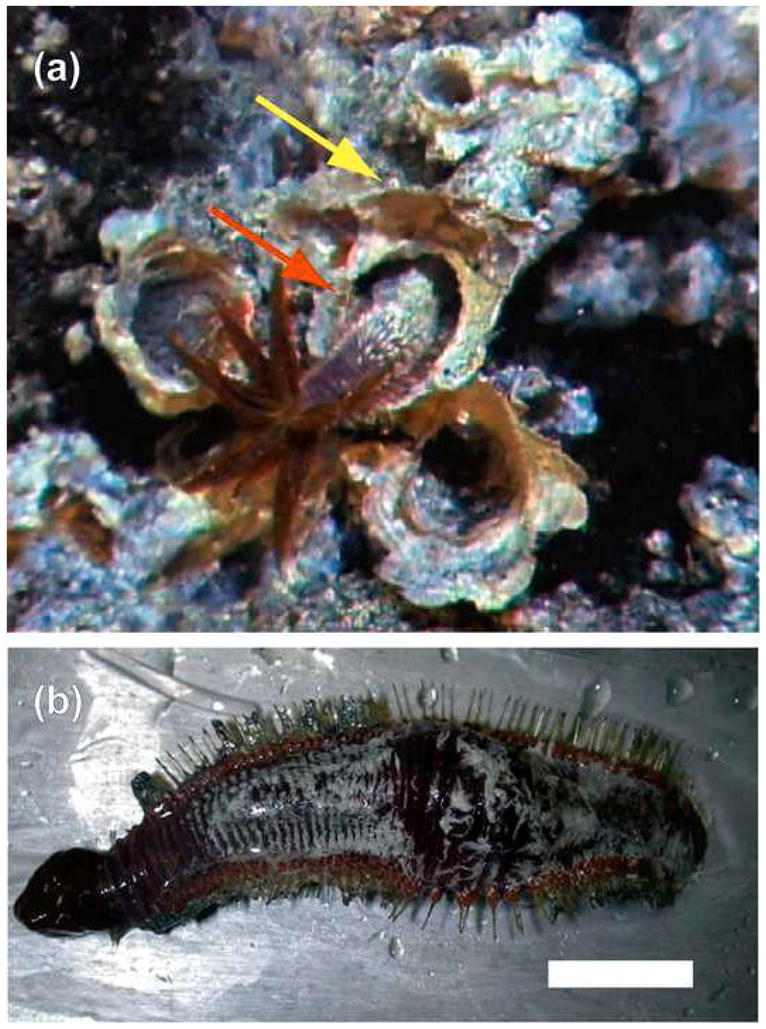
Prokaryotic thermophiles supply stable human protein homologs for structural biology; yet, eukaryotic thermophiles would provide more similar macromolecules plus those missing in microbes. Alvinella pompejana is a deep-sea hydrothermal-vent worm that has been found in temperatures averaging as high as 68 degrees C, with spikes up to 84 degrees C. Here, we used Cu,Zn superoxide dismutase (SOD) to test if this eukaryotic thermophile can provide insights into macromolecular mechanisms and stability by supplying better stable mammalian homologs for structural biology and other biophysical characterizations than those from prokaryotic thermophiles. Identification, cloning, characterization, X-ray scattering (small-angle X-ray scattering, SAXS), and crystal structure determinations show that A. pompejana SOD (ApSOD) is superstable, homologous, and informative. SAXS solution analyses identify the human-like ApSOD dimer. The crystal structure shows the active site at 0.99 A resolution plus anchoring interaction motifs in loops and termini accounting for enhanced stability of ApSOD versus human SOD. Such stabilizing features may reduce movements that promote inappropriate intermolecular interactions, such as amyloid-like filaments found in SOD mutants causing the neurodegenerative disease familial amyotrophic lateral sclerosis or Lou Gehrig's disease. ApSOD further provides the structure of a long-sought SOD product complex at 1.35 A resolution, suggesting a unified inner-sphere mechanism for catalysis involving metal ion movement. Notably, this proposed mechanism resolves apparent paradoxes regarding electron transfer. These results extend knowledge of SOD stability and catalysis and suggest that the eukaryote A. pompejana provides macromolecules highly similar to those from humans, but with enhanced stability more suitable for scientific and medical applications.


Figures

Fig. 1
Fig. 1
Alvinella pompejana. (a) A. pompejana worm (red arrow) projecting out of its vent tube with gills fully extended. The A. pompejana constructed vent tube is noted by the yellow arrow. (b) Ventral view of a ~4 cm Pompeii worm after collection (scale bar = 1 cm) with collapsed gills on the anterior side (red structures on left) and a bulge (middle) from gas expansion due to the ~250 atm change in pressure following collection.
Fig. 2
Fig. 2
ApSOD sequence conservation, fold and residue function. (a) Structure-based alignment of ApSOD with other eukaryotic Cu, Zn SOD proteins with solved structures: Bt, B. taurus; Hs, H. sapiens; Sm, S. mansoni; Xl, X. laevis; So, S. oleracea; Sc, S. cerevisiae. Structural elements and secondary structure of ApSOD are noted above the alignment. Symbols below alignment mark ALS sites in HsSOD: green circle, ApSOD shares wild-type human residue; black square, ApSOD has a different residue; red star, ApSOD residue represents an ALS mutation. Letters below alignment: C, copper-binding ligand; D, disulfide cysteine; B, bridging histidine; Z, zinc-binding ligand; H, H2O2 liganding residue; S, stacking residue, S*, stacking residue hydrogen-bonding partner. Numbers below alignment represent paired interactions noted in Fig. 8 and Table 2. Underlined numbers refer to main-chain atoms involved in interactions, except Glu68, which also makes side-chain interactions. P denotes stabilizing proline caps. (b) Phylogenetic tree for eukaryotic SODs shows estimated evolutionary divergences. Labels at nodes represent bootstrap values and branch lengths are indicated (scale bar = 0.1). (c) Stereoview of the ApSOD structure. Key structural elements in (a) are color coded with abbreviations (VL, Variable loop; GK1 and GK2, Greek key loops 1 and 2; Disulfide region, S-S; ZnBR, Zinc Binding region; EL, Electrostatic loop) otherwise β-strands are cyan and loops are gray. N and C denote termini. The black bar (left) marks the potential dimer interface, on the opposite side of the subunit from the active channel. Metal-liganding residues are shown as sticks with bridging histidine His61 in yellow.
Fig. 3
Fig. 3
ApSOD active site. (a) Stereopair showing trigonal coordination of Cu(I) by histidine ligands in the 0.99 Å ApSOD structure. Composite omit 2Fo-Fc density contoured at 3σ clearly defines individual atoms. Bonding distances with experimentally defined standard uncertainty errors are noted by arrows. Ligand-Cu-ligand angles with errors are shown between respective bonds. (b) Rotation of (a) to show density for the two copper positions. Copper colored spheres are scaled to reflect occupancy. (c-f) Overlay of the 0.99 Å resolution ApSOD model (light green carbon and dark green solvent atoms) and the 1.35 Å resolution ApSOD-H2O2 model (salmon colored carbon and red solvent atoms). (c) Simulated annealing 2Fo-Fc omit electron density (light and dark green) for the solvent in the 0.99 Å non-complexed ApSOD structure and (d) for the 1.35 Å peroxide containing structure (light and dark red). By contouring 2Fo-Fc omit electron density at high levels the center of mass for the constituent in the predicted active site can be located: (c) water W0 and (d) H2O2 contoured at 5σ and 3σ, respectively. (c) Lower level contours of 3σ show a residual curved tail for W0, which suggests that while W0 lies near the hydrogen peroxide O2 position, the water molecule is mobile as it is bound in a site reserved for a larger molecule or that a minor component of the occupancy may reside near the peroxide O1 position. (d) Lower 2σ density for the peroxide soaked structure is cylindrical and conforms to 2 oxygen atoms with realistic hydrogen bonding distances to protein and solvent. Hydrogen bonds are shown as dots. The W0/H2O2 bond to Cu(II) is shown as dashes and bonds between copper and protein as solid lines. (e and f) Rotation of the view in (c and d) to show details of other solvent atoms in the active site. For clarity of position and movements (e) is contoured at 4 and 3σ, and (f) at 3.5 and 2.5σ. (c and e) When water is bound in the active site, W7 is bonded to Arg141, which may play a role in maintaining the active site channel to attract superoxide. (d and f) Arg141 then likely plays a role in catalysis through interactions with the substrates and products.
Fig. 4
Fig. 4
Unified structure-based mechanism for SOD. (a and b) Stereo views of ApSOD active sites from the (a) 0.99 Å structure and (b) the 1.35 Å H2O2 complex structure show alternative solvent positions and bonding, due to replacement of W0 with H2O2, and serve as a guide to the mechanism scheme. Hydrogen bonds are shown as purple balls and bonds to metals are colored matching the metal, where those to protein are solid. (c-g) Scheme for SOD mechanism. Green arrows denote movements and red dashed lines split the upper and lower copper positions. Important atoms in the second half reaction (e-g) are blue. (c and d) First a radical displaces active site Cu(II) binding water W0. Then O2.− hydrogen bonds one oxygen, termed O1, to Arg141 NH1, while the other, termed O2, bonds Cu(II). (d and e) This reduces Cu(II) to Cu(I), shifting it >1 Å deeper into the active site channel, while simultaneously breaking its bond to His61. His61 Nε2 is then protonated, rotating the side chain away from Cu(I) and O2 is released. (e and f) During the second half reaction, a second O2.− is bound within the same site, due to sterics imposed by protein and bonds to His61, Arg141 and W4. To donate a negatively charged electron to the superoxide O2 atom, positively charged Cu(I) is attracted to and moves upward toward O2.− such that orbital overlap occurs for inner sphere electron transfer. His61 Nε2 also donates a proton to O2.− prior to or simultaneously to the electron transfer to form a copper-hydroperoxide intermediate. (f and g) This intermediate is converted to Cu(II) and H2O2 following addition of another proton likely from W4 in trans to avoid steric interference to produce H2O2. After the transfers, Cu(II) is coordinated to His61, and W2 and W3 likely reorient the H2O2 orbitals to that of H2O2 in the liquid state favoring reaction equilibrium toward the product.
Fig. 5
Fig. 5
Thermal anisotropy probability ellipsoids for the ApSOD Cu-binding region. (a) The higher resolution 0.99 Å ApSOD active-site structure. (b) The 1.35 Å ApSOD-H2O2 complex active-site structure. The thermal ellipsoids corresponding to the upper Cu ion position point between the O2.−/H2O2 binding site and W1. The shape of the upper Cu ion ellipsoid from the H2O2 complex structure defines the expected path of Cu ion movement during catalysis (Supplementary Fig. S5). The anisotropic B-factor ellipsoids for the His61 side chain match the motion expected to form a bond with Cu(II) in the upper position and to break the bond to Cu(I) in the lower position.
Fig. 6
Fig. 6
Determination of ApSOD dimer assembly and an ab initio structure in solution by SAXS. (a) Comparison of the experimental SAXS profile for ApSOD (black dots) to scattering profiles calculated from crystal structures of the bacterial SOD dimer from A. pleuropneumoniae (magenta line) and of HsSOD, both as a single subunit (orange line) and dimer (blue line), indicate that ApSOD assembles as a human-like SOD dimer. (b) Comparison of the experimental SAXS profile for ApSOD (black dots) to scattering profiles calculated from the crystallographically defined ApSOD subunit (red line) and dimer (green line) further supports ApSOD dimer assembly. (c) Comparison of the P(r) pairwise interatomic distance functions from ApSOD SAXS data (black line) with P(r) functions calculated for the single subunit (red line) and dimer (green line) crystal structure models confirms dimer assembly in solution. Scattering intensity (ordinate) is plotted against the interatomic pair distances (abscissa). (d and e) X-ray crystal structure of the crystallographic symmetry-related ApSOD dimer model (ribbon diagram) docked into an ab initio SAXS structure solution (surface).
Fig. 7
Fig. 7
ApSOD high stability. Pompeii worm SOD is more stable than human SOD. Circular dichroism indicates that the unfolding midpoint for ApSOD denaturation requires nearly 1 M higher guanidine than the unfolding midpoint for HsSOD.
Fig. 8
Fig. 8
Additional loop and termini interactions may stabilize ApSOD native fold and assembly. (a) Overlay of eukaryotic SOD structures from the 7 species listed in Fig. 2a. Coloring in a spectrum according to relative Cα B-factors distinguishes more flexible, and therefore potentially less stable, parts of the molecule (red, high B-factor) from more rigid regions (dark blue, low B-factor). The most flexible parts are loops and turns localized in 3 areas, designated top, functional and terminal. (b-d) Residues in the 3 most flexible areas of the SOD structure that form stabilizing features in ApSOD that are underrepresented in other structures (see Table 2). Subunits are colored separately. The zinc-binding region is colored the same as the zinc ion in (c) to distinguish it from the electrostatic loop (EL), and the variable loop (VL) is pink in (d).
Fig. 9
Fig. 9
Implications for ALS. (a) Lys21 and Lys98 in ApSOD represent charge reversal ALS mutations E21K and E100K in humans, but are likely not destabilizing in ApSOD due to charge neutralization at the structurally adjacent and intervening position Thr28 (human Lys30). HsSOD is labeled and colored in orange. (b) A stacking arrangement bridges the C-terminus and Greek key loop 2 across the ApSOD dimer interface and continues across the subunit to the zinc-binding region. In ApSOD, unconserved His65 at the end of the stack distal to the dimer interface makes a charged hydrogen or salt bridge bond with unconserved Asp108. The two subunits are colored yellow and teal, and loops within the yellow subunit are colored according to Fig. 2a. A dot surface aids identification of the stacking elements. Dimer symmetry mate residues are labeled with “−B”. (c) The stacking arrangement in HsSOD. The two subunits are colored orange and light brown. Side chains involved in ALS mutations are shown in green on one subunit and magenta on the other. The mutation sites lie at positions that bridge the dimer interface and sandwich the stack together.
Fig. 10
Fig. 10
Implications for amyloid-like filament nucleation and aggregate formation. (a and b) A unified model for nucleation of amyloid-like filaments and formation of soluble aggregates. Due to flexibility in loop and termini regions, over time both (a) ApSOD and (b) HsSOD are likely subjected to reversible local structural perturbations that transiently open structures to expose regions normally sequestered by fold and assembly interactions. ALS SOD mutants that lie within loop and termini regions may cause toxicity by shifting equilibria toward more open forms with decreased barriers to nucleation and growth on non-native, amyloid-like fiber forming contacts. Support for this hypothesis comes from (a) the increased stability of ApSOD, which has additional contacts in these regions, shown as spheres (purple, intrasubunit electrostatic and hydrogen bonding interactions; gray, N-terminal interaction; green, cross-dimer interaction; red, C-terminal and cross-dimer interaction; blue, proline cap; see Fig. 8 and Table 2), that tether these elements and thereby may give ApSOD its higher stability to HsSOD. The interactions shared in HsSOD are also shown as spheres. ALS mutations may enhance local perturbations by removing these and other key contacts, by introducing charge repulsion, or by steric hindrance of normally favorable interactions and packing, resulting in non-native amyloid-like-promoting interactions.

References

    1. Egorova K, Antranikian G. Industrial relevance of thermophilic Archaea. Curr Opin Microbiol. 2005;8:649–55. - PubMed
    1. Yano JK, Poulos TL. New understandings of thermostable and peizostable enzymes. Curr Opin Biotechnol. 2003;14:360–5. - PubMed
    1. Fan L, Williams RS, Shin DS, Chapados BR, Tainer JA. Master keys to DNA replication, repair, and recombination from the structural biology of enzymes from thermophiles. In: Robb FT, Antranikian G, Grogan D, Driessen A, editors. Thermophiles: biology and technology at high temperatures. CRC Press; Boca Raton, FL: 2008. pp. 239–263.
    1. DiDonato M, Deacon AM, Klock HE, McMullan D, Lesley SA. A scaleable and integrated crystallization pipeline applied to mining the Thermotoga maritima proteome. J Struct Funct Genomics. 2004;5:133–46. - PubMed
    1. Robinson-Rechavi M, Alibes A, Godzik A. Contribution of electrostatic interactions, compactness and quaternary structure to protein thermostability: lessons from structural genomics of Thermotoga maritima. J Mol Biol. 2006;356:547–57. - PubMed

Publication types

MeSH terms