. 2009 Dec 8;106(49):20782-7.
doi: 10.1073/pnas.0809510106. Epub 2009 Nov 19.

The synaptic proteins neurexins and neuroligins are widely expressed in the vascular system and contribute to its functions

Affiliations

The synaptic proteins neurexins and neuroligins are widely expressed in the vascular system and contribute to its functions

Alessia Bottos et al. Proc Natl Acad Sci U S A. .

Abstract

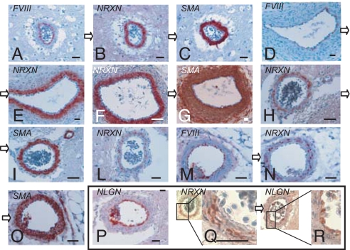
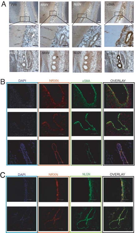
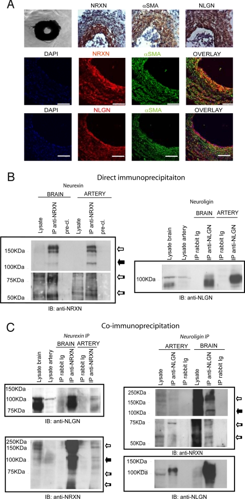
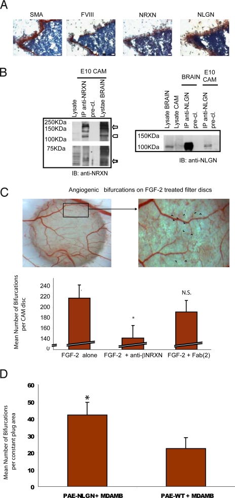
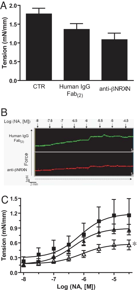
Unlike other neuronal counterparts, primary synaptic proteins are not known to be involved in vascular physiology. Here, we demonstrate that neurexins and neuroligins, which constitute large and complex families of fundamental players in synaptic activity, are produced and processed by endothelial and vascular smooth muscle cells throughout the vasculature. Moreover, they are dynamically regulated during vessel remodeling and form endogenous complexes in large vessels as well as in the brain. We used the chicken chorioallantoic membrane as a system to pursue functional studies and demonstrate that a monoclonal recombinant antibody against beta-neurexin inhibits angiogenesis, whereas exogenous neuroligin has a role in promoting angiogenesis. Finally, as an insight into the mechanism of action of beta-neurexin, we show that the anti-beta-neurexin antibody influences vessel tone in isolated chicken arteries. Our finding strongly supports the idea that even the most complex and plastic events taking place in the nervous system (i.e., synaptic activity) share molecular cues with the vascular system.


Conflict of interest statement

The authors declare no conflict of interest.

Figures

Fig. 1.
Fig. 1.
Neurexin and neuroligin expression in large blood vessels. Pictures show immunostaining of blood vessels from adult chicken brain (A–C, H, I, L, P–R), E11 chicken embryos (D–G), and adult mouse kidneys (M–O). Sections were stained with polyclonal anti-NRXN (B, E, F, H, L, N, Q), anti-NLGN 4F9 (P, R), anti-αSMA (C, G, I, O), or anti-FVIII (A, D, M) antibodies. Sections linked by open arrows (A–C; D and E; F and G; H and I; M–O; Q and R) are consecutive. (Scale bar, 50 μm) See SI Text R1 for a further description of these results.
Fig. 2.
Fig. 2.
Expression and co-localization of neurexin and neuroligin in early (E5) chicken embryo. (A) Immunostaining of E5 chicken embryo sagittal sections showing comparison of endothelial markers, neurexin, neuroligin, and α-SMA. Class 3 vessels (see Table 1) are shown in low-magnification (Top) and high-magnification (Middle) images of a cephalic region proximal to the developing optic lobe. Sections of blood vessels of various wall thicknesses located peripherally to the optic lobe as well as vessels entering the nerve tissues are stained by the neurexin, neuroligin, α-SMA, and FVIII antibodies. (Bottom) Class 4 vessels: aortic arches expressing neurexin, neuroligin, α-SMA, and VEGFR-2. (B) Immunofluorescence and confocal analysis of E5 chicken embryo sagittal sections showing that neurexin (red) is expressed by αSMA-positive cells (green) in 3 examples of developing vessels: a portion of the vessel wall of the developing aorta (Top), an aortic arch section (Middle), and a small vessel of the head vascular plexus (Bottom). (C) Co-localization between neurexin (red) and neuroligin (green) in 2 different vessels: a section of a developing aortic vessel wall (Upper) and a portion of the neck vascular plexus (Lower). DAPI (blue) is nuclear staining. (Scale bar, 50 μm.)
Fig. 3.
Fig. 3.
Neurexin and neuroligin expression pattern, protein production, and co-precipitation in the E18 chicken embryo arteries. (A) (Top) Phase-contrast and immunohistochemical staining of artery sections. (Middle and Bottom) Immunofluorescence and confocal analyses reveal the co-localization of neurexin and neuroligin with αSMA-expressing cells. (Middle) Polyclonal anti-NRXN (red) and anti-αSMA (green) staining. (Bottom) Monoclonal anti-NLGN (red) and anti-αSMA (green) staining. (B and C) E18 chicken embryo arteries were lysed using immunoprecipitation or co-immunoprecipitation buffers (see Materials and Methods) (B) (Left) α and β isoforms of neurexin were immunoprecipitated from both E18 chicken embryo arteries and brain with the polyclonal anti-NRXN. Immunoblotting was performed with monoclonal anti-NRXN. (Right) Neuroligins were immunoprecipitated from E18 chicken embryo arteries and brain with the polyclonal anti-NLGN antibody. Immunoblotting was performed with monoclonal anti-NLGN. (C) Co-immunoprecipitation of neurexin and neuroligin in E18 chicken embryo arteries and brain. (Left) Similar neuroligin isoforms were immunoprecipitated with neurexin in the arteries and brain. (Right) β-Neurexin isoforms preferentially precipitate with neuroligin in chicken embryo arteries, whereas both α- and β-neurexin were enriched by neuroligin in the brain. Immunoblots shown are representative of 3 experiments. White arrow, α-neurexin; black arrow, 115-KDa neurexin; striped arrow, β-neurexin.
Fig. 4.
Fig. 4.
Neurexin and neuroligin expression in the CAM and their role in angiogenesis. (A) Immunohistochemical expression analysis of neurexin and neuroligin in CAM blood vessels. CAMs were sectioned and stained with anti-αSMA, anti-FVIII, polyclonal anti-NRXN, and monoclonal anti-NLGN antibodies. Images show that neurexin and neuroligin are expressed in developing vessels of the E10 chicken CAM. (Scale bar, 50 μm) (B) Biochemical analysis of neurexin and neuroligin expression on E10 CAM: different isoforms of neurexin were immunoprecipitated from the CAM tissue with the polyclonal anti-NRXN antibody (Left). Neuroligins were immunoprecipitated from E10 chicken CAM as in the brain with the polyclonal anti-NLGN antibody (Right). (C) Effect of the recombinant anti-βNRXN antibody on FGF-2–induced angiogenesis. The CAM angiogenesis assay was used to evaluate the effects of the recombinant anti-βNRXN antibody on sprouting angiogenesis. The antibody decreases the number of vessel bifurcations compared with treatment with human IgG Fab(2) or with FGF-2 alone. (Top) A snapshot of the CAM disc. Blue arrowheads indicate the bifurcation used for scoring the result of the graph. (Bottom) Bars represent the number of vessel bifurcations per disc counted after the different treatments. n = 35 for FGF-2 alone, n = 31 for FGF-2 + anti-βNRXN antibody; n = 31 for FGF-2 + human IgG Fab(2). Error bars indicate 95% confidence intervals (CI). ANOVA gave F = 11.081. *, P < 0.01 for anti-βNRXN antibody vs. FGF-2 alone and FGF-2 + human IgG Fab(2) by Student Newman-Keuls test. N.S., P = 0.109 for FGF-2 alone vs. FGF-2 + human IgG Fab(2) by Student Newman-Keuls test. The mean angiogenic level in the untreated CAM discs (not presented in the graph) was 26.5 ± 12% (95% CI) or 26.5 ± 6% (SEM) higher than in the samples treated with anti-β-neurexin (n = 29). (P < 0.01 for anti-βNRXN antibody vs. untreated CAM discs. P = 0.91 for IgG Fab(2) vs. untreated CAM) (D) Pro-angiogenic effect of neuroligin 1 overexpression in ECs. Cultrex plugs (see Materials and Methods) of either nontransfected or neuroligin 1-overexpressing porcine aortic ECs (PAE cells) mixed with the tumor cell line MDA-MB-435 were incubated on CAM for 2 days, and vessel bifurcation was counted as in C. Bars represent the mean count of bifurcations in a fixed area. Error bars indicate 95% CI. n = 24 for PAE NLGN+MDA-MB-435; n = 16 for PAE-WT + MDA-MB-435. *, P < 0.01. Identical results were obtained with 2 different clones of PAE-NLGN.
Fig. 5.
Fig. 5.
Anti-βNRXN antibody inhibited NA-induced contraction on isolated E18 chicken embryo arteries. (A) Anti-βNRXN did not affect K+-induced contraction. Mesenteric arteries were incubated overnight with medium alone (n = 4), 20 μg/mL of human IgG Fab(2) (n = 7), or 20 μg/mL of anti-βNRXN antibody (n = 7). Bars represent mean tension recorded expressed as mN/mm ± SEM. (B) Original representative traces showing the dose–response curve to NA (0.01–100 μM) of 1 artery incubated with 20 μg/mL of human IgG Fab(2) (green) or 20 μg/mL of anti-βNRXN antibody (red). (C) Effect of recombinant anti-βNRXN on the NA dose–response curve. Mesenteric arteries were incubated overnight with (■) medium alone (n = 4), (▴) 20 μg/mL of human IgG Fab(2) (n = 7), or (▵) 20 μg/mL of anti-βNRXN antibody (n = 7). Anti-βNRXN antibody decreased the mean tension value of mesenteric arteries compared with medium alone or human IgG Fab(2). Data are expressed as mean ± SEM. ANOVA gave F = 10.653. *, P < 0.01 for anti-βNRXN antibody vs. medium alone and P = 0.018 vs. human IgG Fab(2) by Bonferroni posttest.

References

    1. Tabuchi K, Sudhof TC. Structure and evolution of neurexin genes: Insight into the mechanism of alternative splicing. Genomics. 2002;79(6):849–859. - PubMed
    1. Bolliger MF, Frei K, Winterhalter KH, Gloor SM. Identification of a novel neuroligin in humans which binds to PSD-95 and has a widespread expression. Biochem J. 2001;356(Pt 2):581–588. - PMC - PubMed
    1. Rissone A, et al. Comparative genome analysis of the neurexin gene family in Danio rerio: Insights into their functions and evolution. Mol Biol Evol. 2007;24(1):236–252. - PubMed
    1. Sudhof TC. α-Latrotoxin and its receptors: Neurexins and CIRL/låtrophilins. Annu Rev Neurosci. 2001;24:933–962. - PubMed
    1. Song JY, Ichtchenko K, Sudhof TC, Brose N. neuroligin 1 is a postsynaptic cell-adhesion molecule of excitatory synapses. Proc Natl Acad Sci USA. 1999;96(3):1100–1105. - PMC - PubMed

Publication types

MeSH terms

Associated data