. 2008 Aug 15:8:139.
doi: 10.1186/1471-2180-8-139.

Inducible Siphoviruses in superficial and deep tissue isolates of Propionibacterium acnes

Affiliations

Inducible Siphoviruses in superficial and deep tissue isolates of Propionibacterium acnes

Rolf Lood et al. BMC Microbiol. .

Abstract

Background: Propionibacterium acnes is a commensal of human skin but is also known to be involved in certain diseases, such as acne vulgaris and infections of orthopaedic implants. Treatment of these conditions is complicated by increased resistance to antibiotics and/or biofilm formation of P. acnes bacteria. P. acnes can be infected by bacteriophages, but until recently little has been known about these viruses. The aim of this study was to identify and characterize inducible phages from P. acnes on a genetic and morphological basis.

Results: More than 70% (65/92) of P. acnes isolates investigated have inducible phages, classified morphologically as Siphoviruses. The phages have a head of 55 nm in diameter and a tail of 145-155 nm in length and 9-10 nm in width. There was no difference in carriage rate of phages between P. acnes isolates from deep infections and isolates from skin. However, there was a significant lower carriage rate of phages in P. acnes biotype IB, mostly attributed to the low carriage rate of inducible phages in biotype IB isolated from deep tissue. Most phages have a strong lytic activity against all P. acnes isolates with inducible phages, but have less lytic activity against isolates that have no prophages. Phages only infected and lysed P. acnes and not other closely related propionibacteria. All phages could infect and lyse their non-induced parental host, indicating that these prophages do not confer superinfection immunity. The phages have identical protein pattern as observed on SDS-PAGE. Finally, sequencing of two phage genes encoding a putative major head protein and an amidase and showed that the phages could be divided into different groups on a genetic basis.

Conclusion: Our findings indicate that temperate phages are common in P. acnes, and that they are a genetically and functionally homogeneous group of Siphoviruses. The phages are specific for P. acnes and do not seem to confer superinfection immunity.


Figures
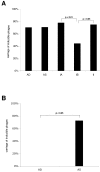
Figure 1
Figure 1
Carriage rate of phages in different groups and biotypes of P. acnes. Phages were induced with 2 μg/ml mitomycin C, lysate sterile filtered and stored for seven days to screen out unstable phages. The lysate was then applied at different concentrations to an overlay plate with the host isolate. If plaques were observed after two days, the sample was regarded positive for phages. (A) A comparison in carriage rate of inducible phages between deep isolates (AD), skin isolates (AS) and biotype IA, IB and II. (B) A comparison of carriage rate of inducible phages in biotype IB between deep isolates and isolates from skin induced by 2 μg/ml mitomycin C.
Figure 2
Figure 2
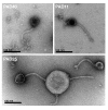
Electron micrographs of bacteriophages from P. acnes. Phages were negatively stained with 0.75% uranyl formate and subjected to transmission electron microscopy. The phages have a head of approximately 55 nm in diameter, loaded with genetic material. Their tails have a size of 150 × 10 nm and are flexible and non-contractile. In the lower micrograph, PAD25 is adhering to bacterial cell debris, and two phages have lost their heads. At the attachment site between the phage and the cell debris, a base plate with attached spikes can be observed. All phages were classified as Siphoviruses based on their morphology.
Figure 3
Figure 3
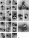
P. acnes bacteriophages classified as Siphoviruses. Phages were negatively stained with 0.75% uranyl formate. All phages were classified as Siphoviruses based on their morphology. No difference in morphology could be observed between the different phages. Several of the phages have empty heads and adhere to bacterial cell debris.
Figure 4
Figure 4
Phylogenetic trees of phages from P. acnes. A gene encoding a putative major head protein and a gene encoding a putative amidase were sequenced in nine P. acnes phages and aligned using MacVector ClustalW alignment. Phylogenetic trees were constructed using neighbor joining with best tree mode. The putative major head protein (A) was similar between all P. acnes phages examined and showed the highest similarity to Mycobacterium phage Che9d gp7, but did also have high similarity to Lactococcus phage phiLC3 MHP and Streptococcus phage SM1 gp40. If outgroups were removed (B) four separate groups of major head proteins could be observed. One group with PA6, another with PAS2 and PAS50, a third group with PAD21 and a forth group with PAS10, PAS12, PAS40, PAD9, PAD20 and PAD42. The putative amidase (C) showed similar patterning among the phages with phages PAS2 and PAS50 representing one group closely related to PA6, while the other phages PAS10, PAS12, PAS40, PAD9, PAD20, PAD21 and PAD42 formed a second group. The closest known phage protein with similarity to the putative amidase is represented by Mycobacterium phage PG1 gp49, but more related is P. acnes own amidase.
Figure 5
Figure 5
P. acnes bacteriophages host-range groups. The host-range for phages isolated from P. acnes was determined by using a bacterial overlay of different P. acnes isolates and adding phages. Four bacterial isolates (KPA171202, AD7, AS1 and AS5) were used to divide the phages into different host-range groups. Phages in host-range group PA I could infect and lyse all four isolates, PA II all except for KPA171202, PA III (AD7, AS1), PA IV (AD7), PA V (AD7, AS5), PA VI (PAD7, PAS42), PA VII (AS1, AS5), PA VIII (AS1, AS5, KPA171202) and PA IX could not infect and lyse any of the isolates used.

References

    1. Webster GF. Acne vulgaris. BMJ. 2002;325:475–479. doi: 10.1136/bmj.325.7362.475. - DOI - PMC - PubMed
    1. Tunney MM, Patrick S, Gorman SP, Nixon JR, Anderson N, Davis RI, Hanna D, Ramage G. Improved detection of infection in hip replacements. A currently underestimated problem. J Bone Joint Surg Br. 1998;80:568–572. doi: 10.1302/0301-620X.80B4.8473. - DOI - PubMed
    1. Zeller V, Ghorbani A, Strady C, Leonard P, Mamoudy P, Desplaces N. Propionibacterium acnes: an agent of prosthetic joint infection and colonization. J Infect. 2007;55:119–124. doi: 10.1016/j.jinf.2007.02.006. - DOI - PubMed
    1. Nord CE, Oprica C. Antibiotic resistance in Propionibacterium acnes. Microbiological and clinical aspects. Anaerobe. 2006;12:207–210. doi: 10.1016/j.anaerobe.2006.08.001. - DOI - PubMed
    1. Ross JI, Snelling AM, Carnegie E, Coates P, Cunliffe WJ, Bettoli V, Tosti G, Katsambas A, Galvan Perez Del Pulgar JI, Rollman O, Torok L, Eady EA, Cove JH. Antibiotic-resistant acne: lessons from Europe. Br J Dermatol. 2003;148:467–478. doi: 10.1046/j.1365-2133.2003.05067.x. - DOI - PubMed

Publication types

MeSH terms

Associated data

LinkOut - more resources