. 2017 Dec 21;12(12):e0190009.
doi: 10.1371/journal.pone.0190009. eCollection 2017.

Unilateral nephrectomy diminishes ischemic acute kidney injury through enhanced perfusion and reduced pro-inflammatory and pro-fibrotic responses

Affiliations

Unilateral nephrectomy diminishes ischemic acute kidney injury through enhanced perfusion and reduced pro-inflammatory and pro-fibrotic responses

Casper Kierulf-Lassen et al. PLoS One. .

Abstract

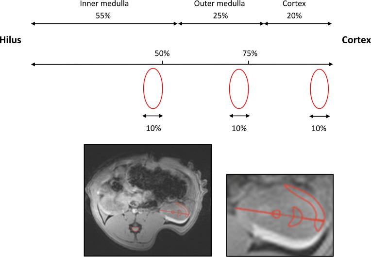
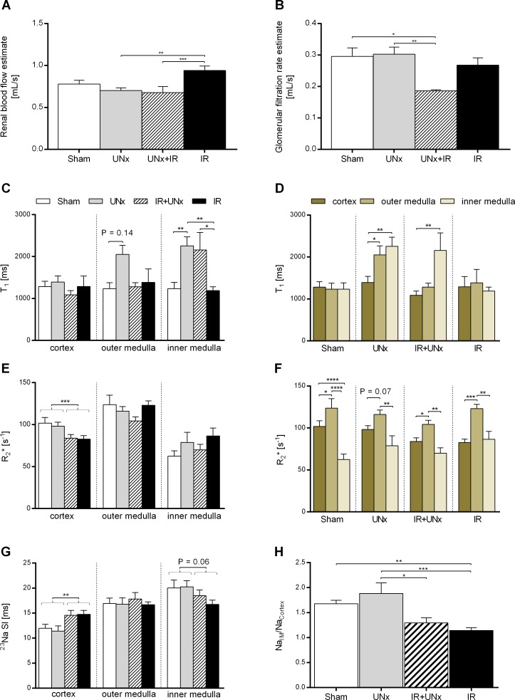
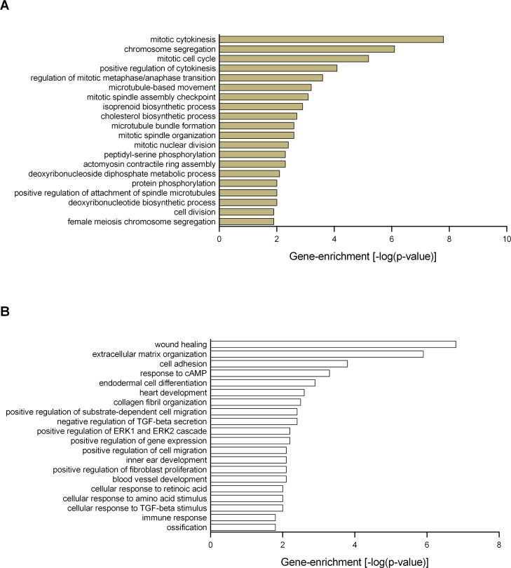
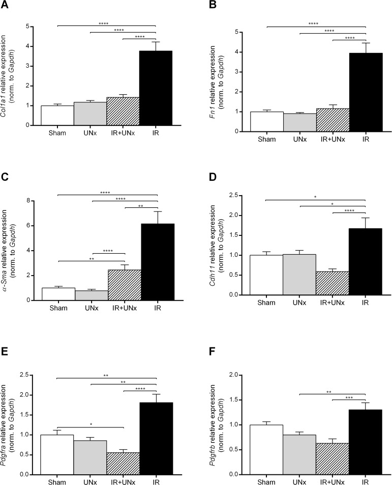
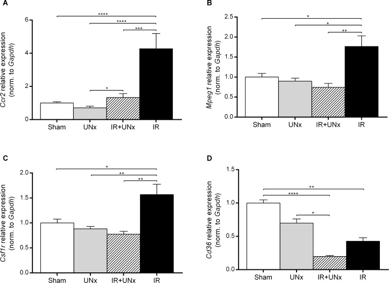
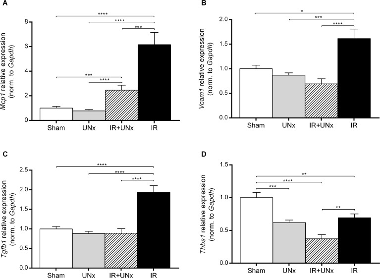
While unilateral nephrectomy (UNx) is suggested to protect against ischemia-reperfusion injury (IRI) in the remaining kidney, the mechanisms underlying this protection remain to be elucidated. In this study, functional MRI was employed in a renal IRI rat model to reveal global and regional changes in renal filtration, perfusion, oxygenation and sodium handling, and microarray and pathway analyses were conducted to identify protective molecular mechanisms. Wistar rats were randomized to either UNx or sham UNx immediately prior to 37 minutes of unilateral renal artery clamping or sham operation under sevoflurane anesthesia. MRI was performed 24 hours after reperfusion. Blood and renal tissue were harvested. RNA was isolated for microarray analysis and QPCR validation of gene expression results. The perfusion (T1 value) was significantly enhanced in the medulla of the post-ischemic kidney following UNx. UNx decreased the expression of fibrogenic genes, i.a. Col1a1, Fn1 and Tgfb1 in the post-ischemic kidney. This was associated with a marked decrease in markers of activated myofibroblasts (Acta2/α-Sma and Cdh11) and macrophages (Ccr2). This was most likely facilitated by down-regulation of Pdgfra, thus inhibiting pericyte-myofibroblast differentiation, chemokine production (Ccl2/Mcp1) and macrophage infiltration. UNx reduced ischemic histopathologic injury. UNx may exert renoprotective effects against IRI through increased perfusion in the renal medulla and alleviation of the acute pro-inflammatory and pro-fibrotic responses possibly through decreased myofibroblast activation. The identified pathways involved may serve as potential therapeutic targets and should be taken into account in experimental models of IRI.


Conflict of interest statement

Competing Interests: The authors have declared that no competing interests exist.

Figures

Fig 1
Fig 1. Regions of interest (ROIs).
The size and position of the cortical, outer and inner medullary ROIs were approximated using the morphometrics of the different renal zones from the study by Oudar et al in male Wistar-Kyoto rats (mean body weight 220 g) [31]. The thicknesses of the zones in percent of the total kidney height along the corticopapillary axis were: cortex = 20%, outer medulla = 25% and inner medulla = 55%. First, the central part of the kidney was localized in the axial anatomical T2-weighted image. A vector was drawn starting from cortex ending at the very tip of the hilus/papilla, covering and measuring the whole kidney height. Two marks were placed in the direction cortex to hilus. The first was set to mark the outer medulla and covered 25% of the total kidney height. The second mark, the inner medulla ROI, was put in the centre and covered 50% of the kidney height. The height of each ROI was set to 10% of the total kidney height. The shape of the different zones on a HE-stained cross-section of the kidney were taken into consideration, when drawing the ROIs. The ROIs, we applied, are to some extent similar to the ones proposed by Oostendorp et al [28].
Fig 2
Fig 2. MRI experiments.
A: Estimates of renal blood flow and B: glomerular filtration rate at 24 hours both adjusted for kidney size. The parameters are based on dynamic contrast enhanced magnetic resonance imaging (DCE-MRI) and analysis using the Patlak-Rutland plot. Intrarenal T1, R2* and 23Na values presented D+F: groupwise and C+E+G: from the different regions of the kidney: the cortex, outer medulla and inner medulla. H: The ratio between sodium signal intensity in inner medulla normalized to cortex, representing the cortico-medullary sodium signal gradient. All values are expressed as mean ± SEM. The effect of UNx and IRI were evaluated by two-way analysis of variance (ANOVA). Group means were compared using two-way ANOVA followed by Bonferroni’s multiple comparisons post-test or the Kruskal-Wallis test with Dunn’s post-test. Means within a group were compared using paired one-way ANOVA or Friedman’s test.* P < 0.05, ** P < 0.01, *** P < 0.005 and **** P < 0.0001. Number of animals: Sham (DCE, n = 7; T1, n = 6; BOLD + 23Na, n = 8), UNx (DCE, n = 7; T1 + BOLD + 23Na, n = 8), IR (n = 8) and IR+UNx (DCE-MRI, n = 5; T1 + BOLD + 23Na, n = 6). IR, ischemia-reperfusion; UNx, unilateral nephrectomy.
Fig 3
Fig 3. Functional enrichment analysis.
The Significance Analysis of Microarrays (SAM) identified A: 56 up-regulated genes (without IRI) and B: 47 down-regulated genes (with IRI) in response to UNx. Functional enrichment analysis was performed using these gene sets in DAVID Bioinformatics Resources, and the enriched Gene Ontology annotations (the biological processes) are presented. The following criteria defined the threshold of an enriched category: a modified Fischer Exact p-value of < 0.05 (> 1.3 after -log10 transformation), and n ≥ 2 genes represented in a category. Number of animals, n = 3 in each group.
Fig 4
Fig 4. Unilateral nephrectomy suppresses genes associated with fibrosis and inflammation.
Pathway analysis was done using Ingenuity ® Pathway Analysis (the diseases and biofunctions application). Unilateral nephrectomy was predicted to inhibit the A: fibrosis and B: inflammation processes following renal ischemia-reperfusion injury by down-regulating the displayed genes. C: Genes involved in both fibrosis and inflammation are identified. Number of animals, n = 3 in each group.
Fig 5
Fig 5. Validation of fibrosis-associated genes.
A: Col1a1, collagen type 1 alpha 1 chain. B: Fn1, fibronectin-1. C: α-Sma, actin, alpha 2, smooth muscle, aorta. D: Cdh11, cadherin-11. E and F: Pdgfra and Pdgfrb, platelet derived growth factor receptor alpha and beta. Bars represent mRNA levels by quantitative polymerase chain reaction in the left renal cortex and outer medulla normalised to Gapdh mRNA. All values are expressed as mean ± SEM. Group means were compared using two-way ANOVA followed by Bonferroni’s multiple comparison post-test. * P < 0.05, ** P < 0.01, *** P < 0.005 and **** P < 0.0001. Number of animals: sham, UNx and IR groups (n = 8), IR+UNx (n = 6). IR, ischemia-reperfusion; UNx, unilateral nephrectomy.
Fig 6
Fig 6. Validation of macrophage expressed genes.
A: Ccr2, C-C chemokine receptor type 2. B: Mpeg1, macrophage expressed 1. C: Csf1r, colony stimulating factor 1 receptor. D: Cd36, cd36 molecule. Bars represent mRNA levels by quantitative polymerase chain reaction in the left renal cortex and outer medulla normalised to Gapdh mRNA. All values are expressed as mean ± SEM. Group means were compared using two-way ANOVA followed by Bonferroni’s multiple comparison post-test. * P < 0.05, ** P < 0.01, *** P < 0.005 and **** P < 0.0001. Number of animals: sham, UNx and IR groups (n = 8), IR+UNx (n = 6). IR, ischemia-reperfusion; UNx, unilateral nephrectomy.
Fig 7
Fig 7. Validation of inflammatory mediators genes.
A: Mcp1, monocyte chemoattractant protein-1. B: Vcam1, vascular cell adhesion molecule-1. C: Tgfb1, transforming growth factor-β1. D: Thbs1, thrombospondin-1. Bars represent mRNA levels by quantitative polymerase chain reaction in the left renal cortex and outer medulla normalised to Gapdh mRNA. All values are expressed as mean ± SEM. Group means were compared using two-way ANOVA followed by Bonferroni’s multiple comparison post-test. * P < 0.05, ** P < 0.01, *** P < 0.005 and **** P < 0.0001. Number of animals: sham, UNx and IR groups (n = 8), IR+UNx (n = 6). IR, ischemia-reperfusion; UNx, unilateral nephrectomy.

References

    1. Sharfuddin AA, Weisbord SD, Palevsky PM, Molitoris BA. Acute Kidney Injury In: Skorecki K, Chertow GM, Marsden PA, Taal MW, Yu ASL, editors. Brenner and Rector's The Kidney. E-Book. 10th ed.: Elsevier Health Sciences; 2016. p. 958–1011.
    1. Nakajima T, Miyaji T, Kato A, Ikegaya N, Yamamoto T, Hishida A. Uninephrectomy reduces apoptotic cell death and enhances renal tubular cell regeneration in ischemic ARF in rats. Am J Physiol 1996. October;271(4 Pt 2):F846–53. - PubMed
    1. Fried TA, Hishida A, Barnes JL, Stein JH. Ischemic acute renal failure in the rat: protective effect of uninephrectomy. Am J Physiol 1984. October;247(4 Pt 2):F568–74. - PubMed
    1. Lin SY, Humphreys MH. Centrally administered naloxone blocks reflex natriuresis after acute unilateral nephrectomy. Am J Physiol 1985. September;249(3 Pt 2):F390–5. - PubMed
    1. Krohn AG, Peng BB, Antell HI, Stein S, Waterhouse K. Compensatory renal hypertrophy: the role of immediate vascular changes in its production. J Urol 1970. May;103(5):564–568. - PubMed

Publication types