. 2008 Aug 7;454(7205):717-21.
doi: 10.1038/nature07219.

Secreted transcription factor controls Mycobacterium tuberculosis virulence

Affiliations

Secreted transcription factor controls Mycobacterium tuberculosis virulence

Sridharan Raghavan et al. Nature. .

Abstract

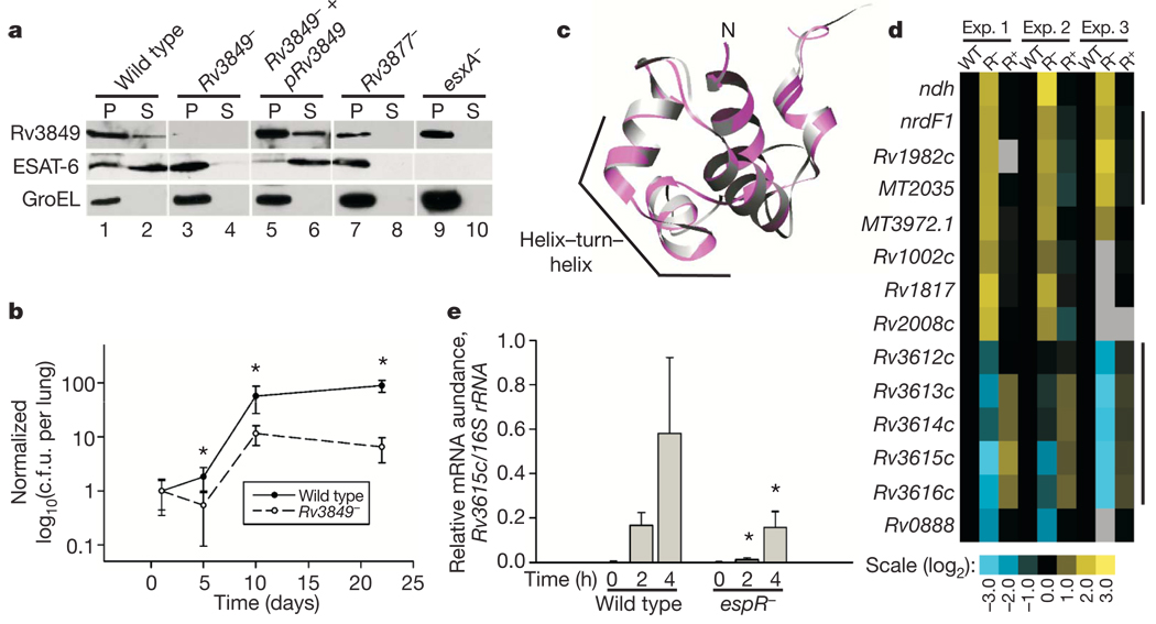
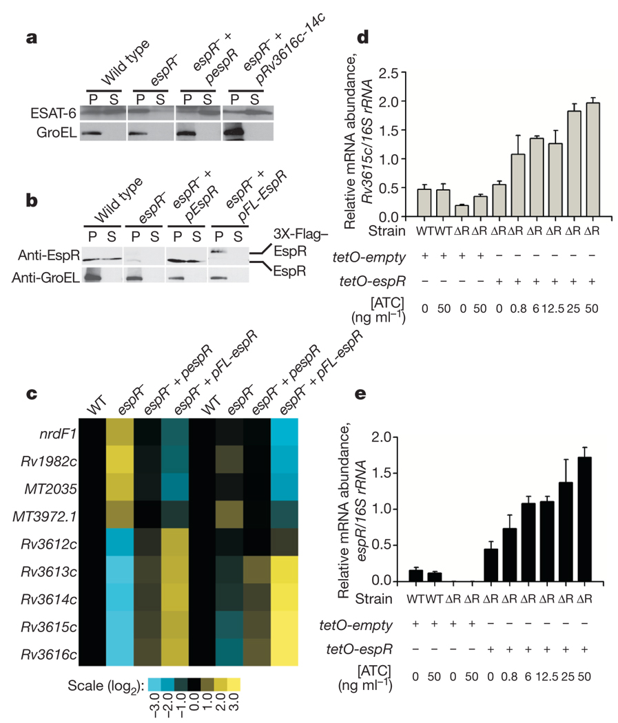
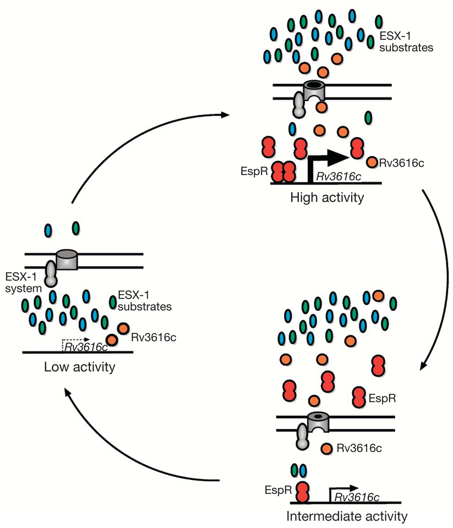
Bacterial pathogens trigger specialized virulence factor secretion systems on encountering host cells. The ESX-1 protein secretion system of Mycobacterium tuberculosis-the causative agent of the human disease tuberculosis-delivers bacterial proteins into host cells during infection and is critical for virulence, but how it is regulated is unknown. Here we show that EspR (also known as Rv3849) is a key regulator of ESX-1 that is required for secretion and virulence in mice. EspR activates transcription of an operon that includes three ESX-1 components, Rv3616c-Rv3614c, whose expression in turn promotes secretion of ESX-1 substrates. EspR directly binds to and activates the Rv3616c-Rv3614c promoter and, unexpectedly, is itself secreted from the bacterial cell by the ESX-1 system that it regulates. Efflux of the DNA-binding regulator results in reduced Rv3616c-Rv3614c transcription, and thus reduced ESX-1 secretion. Our results reveal a direct negative feedback loop that regulates the activity of a secretion system essential for virulence. As the virulence factors secreted by the ESX-1 system are highly antigenic, fine control of secretion may be critical to successful infection.


Figures

Figure 1
Figure 1. Rv3849 is a secreted regulator of the ESX-1 system
a, Cell pellets (P) and culture supernatants (S) from wild-type, Rv3849, Rv3849 complemented, Rv3877, and esxA bacteria were probed for Rv3849, ESAT-6 and GroEL by western blot. GroEL, an intracellular protein used as a lysis control, was detected in cell pellets of all strains and was absent from supernatants. b, C57BL/6 mice were infected with 1 × 10⁶ colony-forming units (c.f.u.) of wild-type and Rv3849 mutant M. tuberculosis through the intravenous route. Bacterial growth was monitored by counting c.f.u. from lung homogenates, and values at each time point were normalized to the initial dose at 24 h post-infection. For each bacterial strain, mean ± s.d. is shown from five mice at each time point. *P < 0.05 between Rv3849 and wild-type. c, The crystal structure of SinR (violet) was used as a template to generate a structural model of Rv3849 (silver) using the program Modeller. The overlay of the N-terminal HTH domains of both proteins is shown. d, Genome-wide transcriptional profiles were examined using M. tuberculosis-specific oligonucleotide microarrays. Gene expression values in Rv3849 (R) and complemented Rv3849 (R+) bacteria were divided by expression values in wild-type (WT) bacteria. Results from three independent experiments are shown. Genes with statistically significant changes in expression are shown (see Supplementary Methods for details of statistical analysis). Black, no change; yellow, increased expression; blue, decreased expression relative to wild-type; grey, missing data. Lines to the right of the cluster diagram represent probable operons. e, Expression of Rv3615c in wild-type and espR bacteria in liquid culture (0 h) and 2 h and 4 h following infection of bone-marrow-derived macrophages was measured by quantitative PCR from amplified total M. tuberculosis RNA. Rv3615c expression was normalized to expression of 16S rRNA. Shown are mean ± s.d. of triplicate measurements from one of 2 experiments. *P < 0.05 comparing wild type and espR at 2 h and at 4 h.
Figure 2
Figure 2. EspR binds and activates the Rv3616c–Rv3612c promoter
a, M. tuberculosis EspR was expressed in M. smegmatis strains carrying lacZ reporter constructs with different length Rv3616c promoter fragments. Activity was monitored by plating strains on media containing X-gal (5-bromo-4-chloro-3-indolyl-β-D-galactoside) and by quantitative β-galactosidase assay. Shown are mean + s.d. of triplicate measurements from one of 3 experiments. b, Binding of EspR to the Rv3616c promoter was assayed by EMSA using purified MBP–EspR fusion protein, or MBP alone, and radiolabelled DNA fragments. c, N-terminal sequences of EspR homologues of other actinomycetes were aligned and the highly conserved residues targeted for mutagenesis are highlighted in red (top). A C-terminal deletion series of EspR was also generated (bottom). d, Secretion of ESAT-6 from espR bacteria expressing mutant forms of EspR was assayed by western blot analysis of cell pellets (P) and culture supernatants (S). e, Expression of the EspR regulon in the espR mutant (R) expressing either wild-type espR (R+) or the indicated mutants was monitored by microarray. Gene expression in each strain was divided by expression level in wild-type (WT) bacteria. Black, no change; yellow, increased expression; blue, decreased expression relative to wild type; grey, missing data.
Figure 3
Figure 3. Examining the role of EspR secretion in ESX-1 function
a, Rv3616c–Rv3614c were expressed in espR bacteria under the control of the constitutive GroEL promoter. ESAT-6 and GroEL were analysed by western blot in cell pellets (P) and culture supernatants (S) of wild-type bacteria, espR bacteria, complemented espR bacteria, and espR bacteria constitutively expressing Rv3616c–Rv3614c. b, Cell pellets (P) and culture supernatants (S) of wild-type bacteria and espR bacteria expressing N-terminally Flag-tagged EspR were probed by western blot for EspR and GroEL. Arrows indicate endogenous EspR and 3 ×-Flag–EspR. c, Microarray analysis comparing expression of EspR regulon genes in the strains used in b. Black, no change; yellow, increased expression; blue, decreased expression relative to wild-type; grey, missing data. Two replicate experiments are shown. d, e, EspR was placed under the transcriptional control of the Tet-inducible promoter and introduced on an integrating plasmid into wild-type (WT) and ΔespR (ΔR) bacteria. Total RNA was harvested from mid-log phase M.tuberculosis grownin the presenceof the indicated concentration of anhydrous tetracycline (ATC). Rv3615c (d) and espR (e) expression were measured by quantitative real-time PCR and normalized to 16S rRNA expression. Shown are mean + s.d. of triplicate measurements from one representative experiment of three.
Figure 4
Figure 4. Model of EspR regulated activation of ESX-1 secretion and negative feedback through export via the ESX-1 pathway
ESX-1 function cycles between low-activity and high-activity states (left and top right, respectively) and is activated by EspR transcriptional activity but limited by EspR secretion. Activation of EspR by unknown stimuli leads to increased transcription (thick black arrow) expression of Rv3616c (orange circles) and other genes in the operon (not shown). Rv3616c accumulation leads to high activity of ESX-1, perhaps via interactions with membrane-bound ESX-1 secretion components (grey), leading to secretion of substrates, including Rv3616c. Secretion of EspR decreases the intracellular concentration of the activator and subsequently lowers Rv3616c transcription, depicted here (thin arrow, bottom right) as an intermediate-activity state. Eventually, this negative feedback loop returns the ESX-1 system back to the low-activity state transcription (dashed arrow).

Comment in

References

    1. Stanley SA, Raghavan S, Hwang WW, Cox JS. Acute infection and macrophage subversion by Mycobacterium tuberculosis require a specialized secretion system. Proc. Natl Acad. Sci. USA. 2003;100:13001–13006. - PMC - PubMed
    1. Hsu T, et al. The primary mechanism of attenuation of bacillus Calmette-Guerin is a loss of secreted lytic function required for invasion of lung interstitial tissue. Proc. Natl Acad. Sci. USA. 2003;100:12420–12425. - PMC - PubMed
    1. Pathak SK, et al. Direct extracellular interaction between the early secreted antigen ESAT-6 of Mycobacterium tuberculosis and TLR2 inhibits TLR signaling in macrophages. Nature Immunol. 2007;8:610–618. - PubMed
    1. Abdallah AM, et al. Type VII secretion-mycobacteria show the way. Nature Rev. Microbiol. 2007;5:883–891. - PubMed
    1. Guinn KM, et al. Individual RD1-region genes are required for export of ESAT-6/ CFP-10 and for virulence of Mycobacterium tuberculosis. Mol. Microbiol. 2004;51:359–370. - PMC - PubMed

Publication types

MeSH terms

Associated data